
Parts
Copyright 2010
Niharika Nixit MD Ashley Davidoff MD
The uterus is divided in to two parts by a constriction called isthmus midway between apex and base. The portion above the isthmus is called as corpus or body of uterus and below the isthmus is called the cervix. The isthmus corresponds to internal orifice of uterus. The portion of body above opening of fallopian tubes is called as fundus.
Corpus Uteri ( Body ) gradually narrows from fundus to isthmus
Vesical or anterior surface is flattened and covererd by peritoneum which reflected on to the bladder to for, vesicouterine space.
Intestinal or posterior surface Is convex transcversely and is covered by peritoneumdown to the cervix and vagina it is in relation with sigmoid colon separated by loops of intestine this is called a pouch of Douglas; Pelvic collections ususlly collect in this space and can be drained vaginally.
Supports of uterus: Uterus remains in its pelvic position by virtue of its positions and various ligaments that keep it in position. These supports can lose strength with multiple vaginal childbirths, menopause and age and cause utero-vaginal position.
|
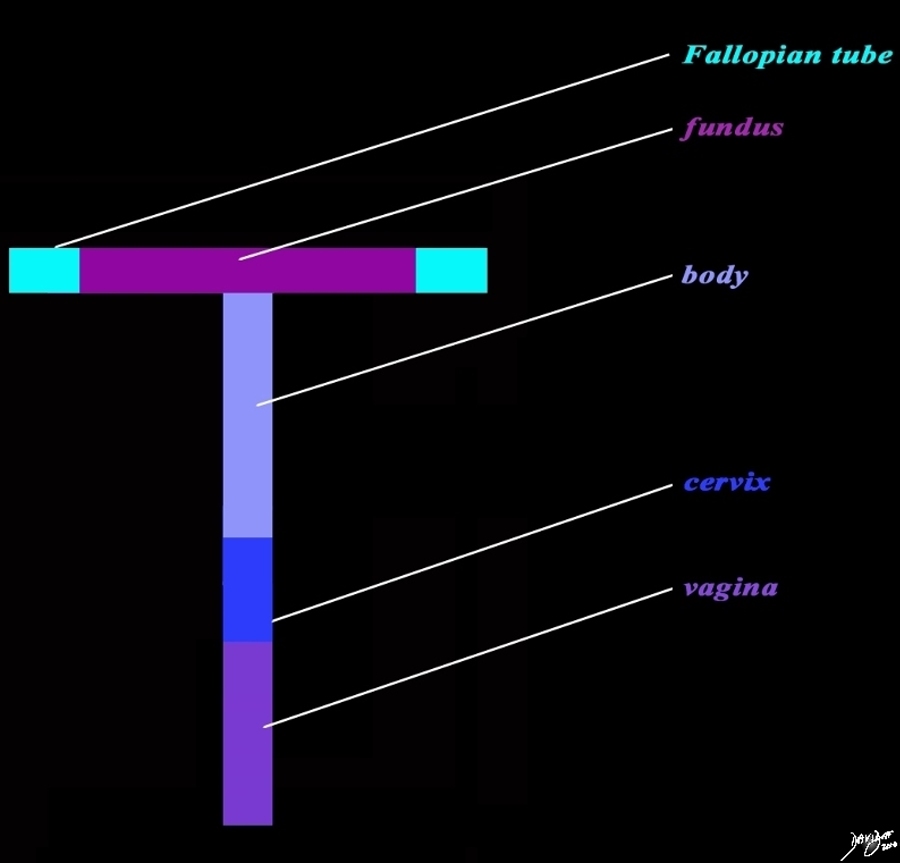
The “T” shaped Uterus and its Component Parts |
|
The diagram reflects the “T” shaped structure of the uterus as well as the component parts including the fundus (dark pink) the body (light purple), and cervix (royal blue). The Fallopian tubes (teal) at the upstream end, and the vagina at the downstream end (purple) complete the tract of the genital tract . Courtesy Ashley Davidoff MD copyright 2010 all rights reserved 96266b02L02h.91s |
|
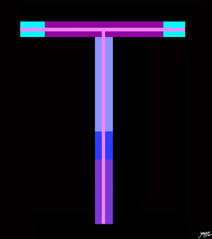
The Tract Within the Component Structures |
|
The diagram reflects the “T” shaped structure of the uterus, the component parts, and the tract (light pink) starting from the vagina (purple and extending through the uterus and Fallopian tubes (teal). The parts that the tract traverses includes the vagina at the downstream end (purple), cervix (royal blue), lower uterine segment (light blue), the body (light purple), the fundus (dark pink) and finally the Fallopian tubes (teal) at the upstream end. Courtesy Ashley Davidoff MD copyright 2010 all rights reserved 96266b03.8sh |
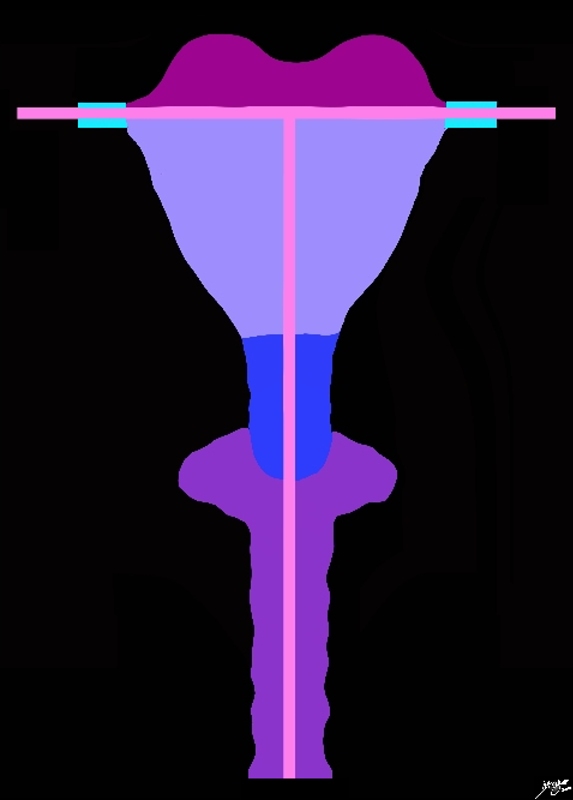
Creating Form around the “T” |
|
The diagram reflects the “T” shaped structure of the uterus, the component parts, and the tract (light pink) starting from the vagina (purple and extending through the uterus and Fallopian tubes (teal). The form of the structures surrounding the tract have been expanded to their relative dimensions and shapes The parts that the tract traverses includes the vagina at the downstream end (purple), cervix (royal blue), lower uterine segment (light blue), the body (light purple), the fundus (dark pink) and finally the Fallopian tubes (teal) at the upstream end. . Courtesy Ashley Davidoff MD copyright 2010 all rights reserved 96266b12bh.81s |
|
Shaping the “T” to Realistic Form |
|
The diagram advances the “T” shaped structure of the uterus and the genital tract. The form of the structures and the tract have been expanded to their relative dimensions and shapes The parts that the tract traverses includes the vagina at the downstream end which now is a significantly expanded part of the tract (pink), cervix (royal blue), lower uterine segment (light blue), the body (lighter blue), the fundus (dark pink) and finally the Fallopian tubes (teal) at the upstream end. Courtesy Ashley Davidoff MD copyright 2010 all rights reserved 96266b15b01h02.8s |
In the Sagittal Plane

“L” Shpaed Structure in the Sagittal |
|
The diagram represents a sagittal view of the uterus reflecting an ‘L” shaped structure of the uterus and vagina The parts that the “L” represents include the the fundus (dark pink), the body (lighter blue), lower uterine segment (light blue), cervix (royal blue), and the vagina (purple) at the downstream end. Courtesy Ashley Davidoff MD copyright 2010 all rights reserved 96267b03h.8s |

The “L”Shaped Uterus and Vagina with the Internal Tract |
|
The diagram represents a sagittal view of the uterus reflecting an ‘L” shaped structure of the uterus vagina, and the internal cavity. The parts that the tract traverses includes the vagina at the downstream end which now is a significantly (pink), cervix (royal blue), lower uterine segment (light blue), the body (lighter blue), and the fundus (dark pink). Courtesy Ashley Davidoff MD copyright 2010 all rights reserved 96267b04h.8s |
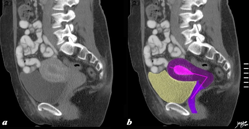 The “L” Shaped Uterus The “L” Shaped Uterus |
| The sagittaly reconstructed CT shows an anteverted uterus buoyed and cushioned by partly filled bladder (yellow) In this sagittal view of the uterus an ‘L” shaped structure of the uterus vagina, and the internal cavity is diagrammed in by the pink vectors of the tract.
Courtesy Ashley Davidoff MD copyright 2010 all rights reserved 60385c04b.8s |
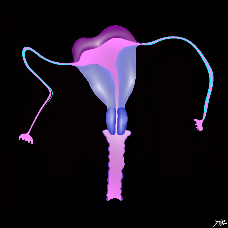
 The Flow of the Fallopian Tubes The Flow of the Fallopian Tubes |
|
46701b03.49k.8s uterus fallopian tubes cervix ovaries broad ligament normal anatomy vagina gravid vertex presentation brain placenta foot rawMRI T2 weighted Courtesy Ashley Davidoff MD Davidoff art copyright 2009 all rights reserved |
 Fallopian Tubes – Connection to Life Fallopian Tubes – Connection to Life |
|
46376b01b01b05 pelvis uterus Fallopian tubes fimbrae uterus uterine cavity cervix vagina fornix hysterosalpingogram Davidoff MD |