Copyright 2010
Introduction
The Enlarged Uterus of Pregnancy
|
Non Gravid , 32 Week Pregnancy, and PostPartum Uterus |
|
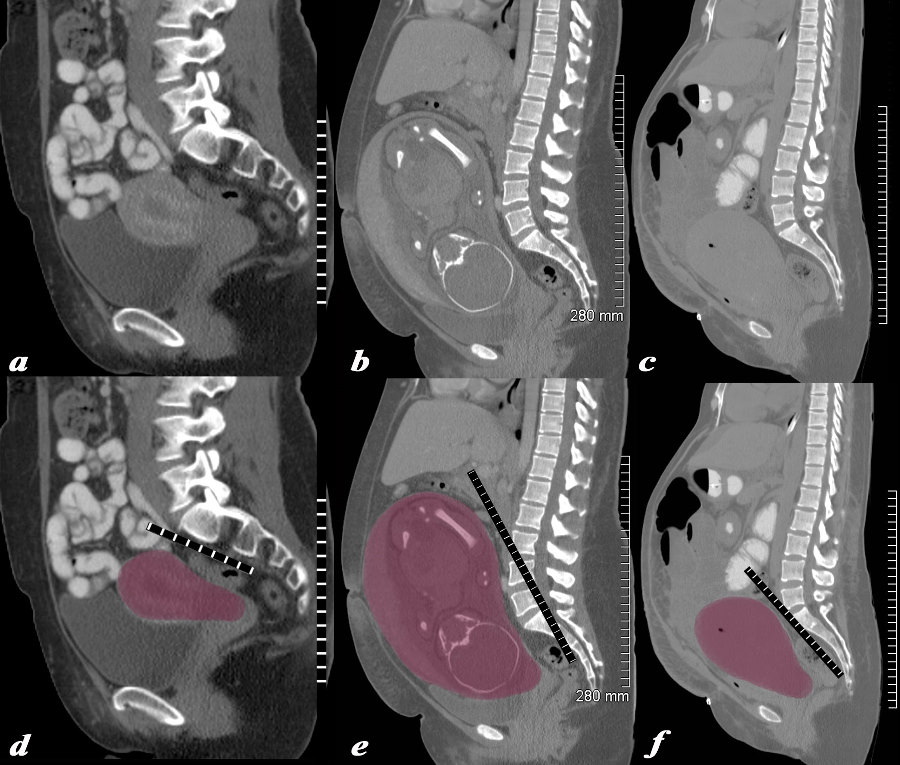
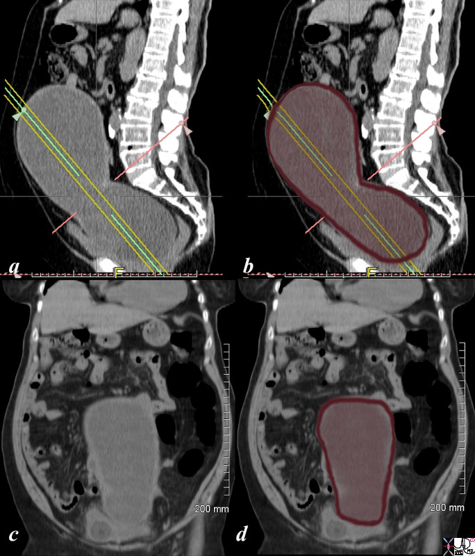
The series of CT scans from different patients are reconstructed in the sagittal plane to show the mature uterus in the non-gravid state (a,d) with a 32 week pregnancy (b,e), and in the postpartum, post cesarean section state (c,f). In the nongravid adult the uterus the craniocaudad span (c-c) measures about 9cms and the anteroposterior (A-P) dimension it measures 4.5cms The uterus containing the 32week pregnancy measures 24cms (c-c) by 16cms (A-P). In the post cesarean section patient the uterus measures 17cms (c-c) by 9cms (A-P). Courtesy Ashley Davidoff MD Copyright 2010 All rights reserved 78093c08.8s |
|
32 week Pregnancy |
|
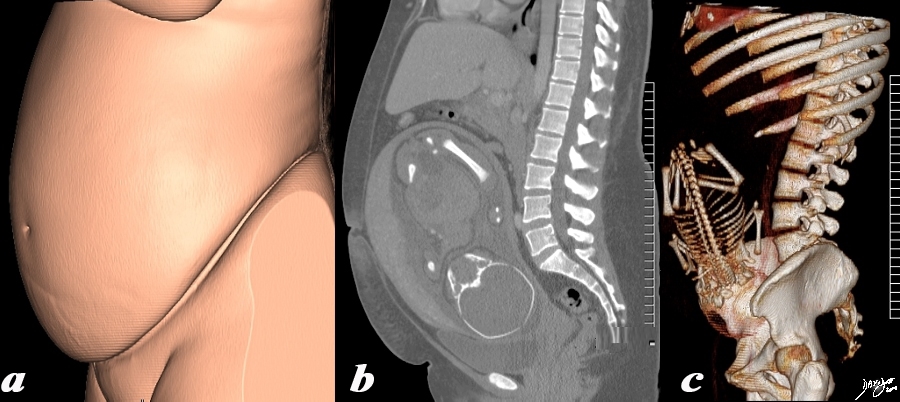
The 3 sagittally reconstructed images of the gravid uterus are from a normal 31 year woman carrying a 32 week gestation. They depict the outside appearance of the patient (a), 2 D sagittal view (b) and 3D sagittal view(c). Pregnancy is the main reason for living for the uterus and the opportunity to use it for this purpose is only a brief time in the long life of an adult female – but the species depends on this brief sojourn. Courtesy Ashley Davidoff MD Copyright 2010 All rights reserved 96332c.91s |
|
Twin Pregnancy – Pushing the Limits |
|
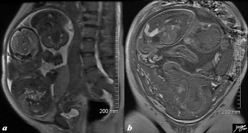
The T2 weighted MRI from a 44 year old patient with 32 week twin gestation in the sagittal plane (a) and in the coronal plane (b) revealing a uterus that measures 30cms in craniocaudad span, by 18cms A-P, by 26cms in the transverse plane. Courtesy Ashley Davidoff MD Copyright 2010 All rights reserved 88845c01.8s |
The Most Common Pathology Causing Enlarged Uterus |
|
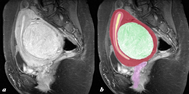
The sagittal SPIR sequence from an MRI study shows a single large fibroid situated in the body of the uterus (green) and displacing and enlarging the remaining uterus forward and upward. The endometrial cavity is compressed (yellow) as is the junctional zone (salmon red) and the myometrium itself (maroon) The uterus has been lifted out of the pelvis and could be palpated clinically. The morphology of the swirling like shape to the myocardial tissue – an exemplary finding under the microscope is well appreciated in this image Courtesy Philips Medical Systems Images rendered by Ashley Davidoff MD Copyright 2010 All rights reserved 96932c01.8s |
Fibroid Uterus
Enlarged and Lobular |
|
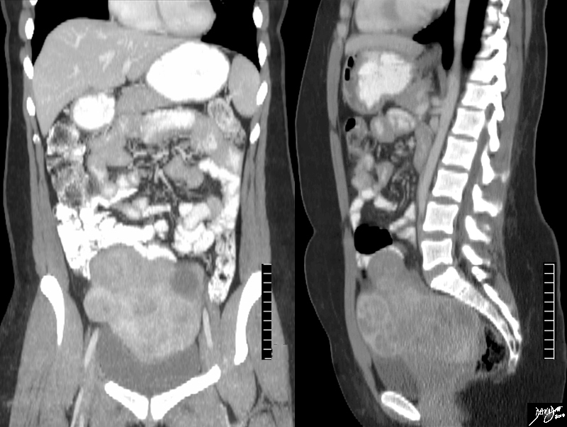
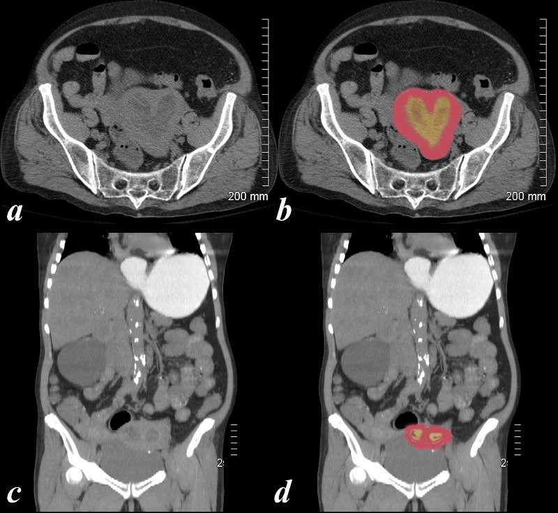
The CTscan is from a 31year old female, who presents with a pelvic mass.The study reveals a large uterus with multicentric fibroids and mild hydronephrosis The heterogeneous and lobular mass measures 17cms (c-c)by 10cms (A-P) by 12cms (transverse), and is consistent with a fibroid Courtesy Ashley Davidoff MD copyright 2009 all rights reserved 84362c.8s |
Adenomyosis

Adenomyosis |
|
This T2 weighted MRI of a 41 year old female shows thickened junctional zone of the uterus measuring up to 12 mms characteristic of adenomyosis . Courtesy Ashey Davidoff MD copyright 2009 14707c01.8s |
Carcinoma
|
Large Uterus Endometrial Carcinoma with Hydrosalpinx |
|
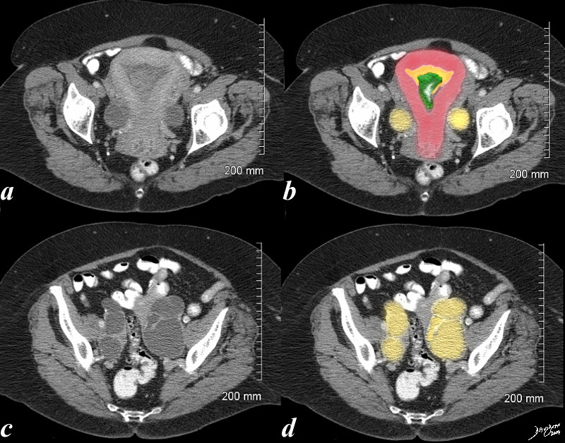
This 70 year old female presents with pelvic discomfort. The CT shows an endometrium (orange in b) filled with complex soft tissue in the endometrial cavity (green in b) and the Fallopian tubes are distended with fluid (yellow in b and d) caused by the obstructing carcinoma. The uterus is enlarged Courtesy Ashley Davidoff MD copyright 2009 all rights reserved 48414c02.8s |
Cervical Carcinoma and Obstruction |
|
46 year old female with obstructed uterus and fluid filled endometrial cavity caused by cervical carcinoma complicated by obstruction. Courtesy Ashley Davidoff MD Copyright 2009 all rights reserved 83648c02.8s |
Carcinoma of the Cervix in a Bicornuate Uterus Complicated by Obstruction |
| 74 year old female with bicornuate uterus and dilated endometrial cavities Diagnosis is carcinoma of the cervix with obstruction . The myometrium is overlaid in dark pink, and the endometrial cavity is a heterogeneous orange consisting of both fluid and soft tissue elements.
Incidental note is made of gastroesophageal reflux Courtesy Ashley Davidoff MD copyright 2009 all rights reserved 83438c01.8s |
Rapidly Enlarging Mass in the Pelvis
|
Leiomyosarcoma |
|
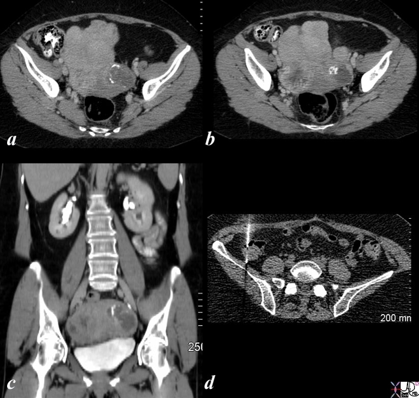
The CT is from a 58 year old post menopausal female who presents with an enlarging pelvic mass (a,b,c). CTscan shows multiple large masses in the uterus some of which are calcified consistent with leiomyomas. However clinically the mass had grown significantly in the last few months. Surgery revealed a leiomyosarcoma. Follow up CT 1 year later showed paracolic nodules which were proven to be metastatic deposits by biopsy (d). Courtesy Ashley Davidoff MD copyright 2009 all rights reserved 85519cL.8s |
Different Criteria for “Large” in the Elderly
|
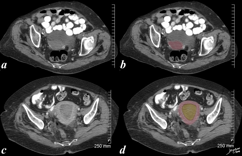
Assessing the Large Uterus in the Elderly Normal (a,b) and Large (c,d) |
|
The first CTscan (a,b) is from an 86 year old patient with a small but normal post menopausal uterus showing characteristic vascular calcification. The second CT (c,d) is from an 83F year old patient with distended endometrial cavity (orange in d) with known uterine carcinoma Courtesy Ashley Davidoff MD copyright 2009 all rights reserved 83731c01.8S |