
The Common Vein Copyright 2010
Definition
The cervix is the lower constricted segment of the uterus. It is somewhat conical in shape with its truncated apex directed downwards and backwards.The cervix projects through the anterior wall of vagina which divides it in to supravaginal and vaginal portion.
Supravaginal portion is related to the bladder anteriorly separated by fibrous tissue called parametrium this extends laterally and uterine arteries enter the cevix through it. The ureters run downward and forward in it about 2 cms from form margin of cervix.
Vaginal portio ( portio vaginalis) Projects free in to the vagina through thte anterior wall dividing it an to anterior and posterior fornices. external orifice on its tip is circular aperture bounded by anterior and posterior lip.
Cavity of the uterus is a triangular shaped ,slit like and flattened antero-posteriorly. base of the triangle is formed by the internal surface of fundus and apex by internal orifice.
cavity of the cervix is a fusiform broader in the middle canal like which communicates with uterine cavity through internal orifice and with vagina through external orifice. It has a posterior and anteriot ridge from which mucous is thrown in to palmate folds giving it a tree like appearance in cross section this is called as arbor Vitae uterina.
Pathway to Conception |
|
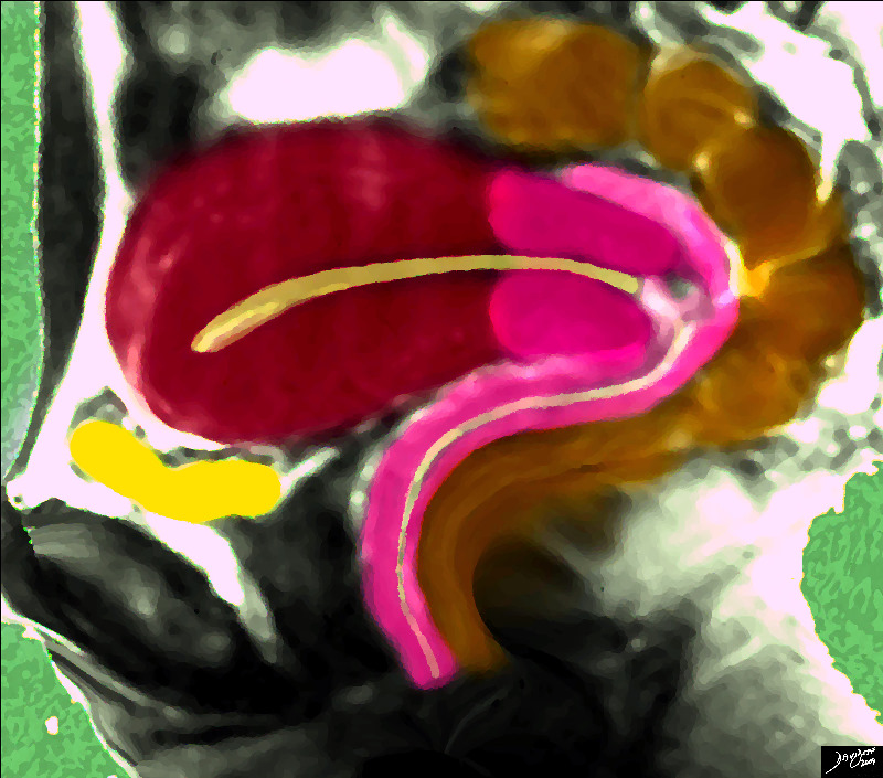
The sagittal diagram of the uterus shows the uterus surrounded by its neighbours, including the empty bladder (yellow) that lies anteriorly and inferiorly and the rectosigmoid colon that lies superiorly and posteriorly. Courtesy Ashley Davidoff MD copyright 2009 14707.2kb04i06.s.4k.8s |
The Internal Cervical Os |
|
Courtesy Ashley Davidoff MD copyright 2009 all rights reserved 46701b03.49k.8s |
Normal PAP smear |
|
Normal PAP smear Courtesy Laura Miller MD 96933B.8 |
Abnormal PAP Smear – Cervical Carcinoma |
|
Abnormal PAP smear Cervical Carcinoma 96933B.8 Courtesy Laura Miller MD 96934b.8 |
The Cervix and the Cervical Canal in Sagittal Projection |
|
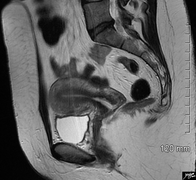
The sagittal STIR sequence from an MRI study shows the normal appearance of the uterus in a 34 year old female. The pear shaped form is exemplified together with the 3 parts of the uterus. The inner endometrium of intermediate signal, the junctional zone of low signal consisting of compacted smooth muscle with low water content, and the surrounding myometrium of smooth muscle with a higher water content. Courtesy Ashley Davidoff MD Copyright 2010 All rights reserved 96785.8s |
Axial Plane Coronal Projection |
|
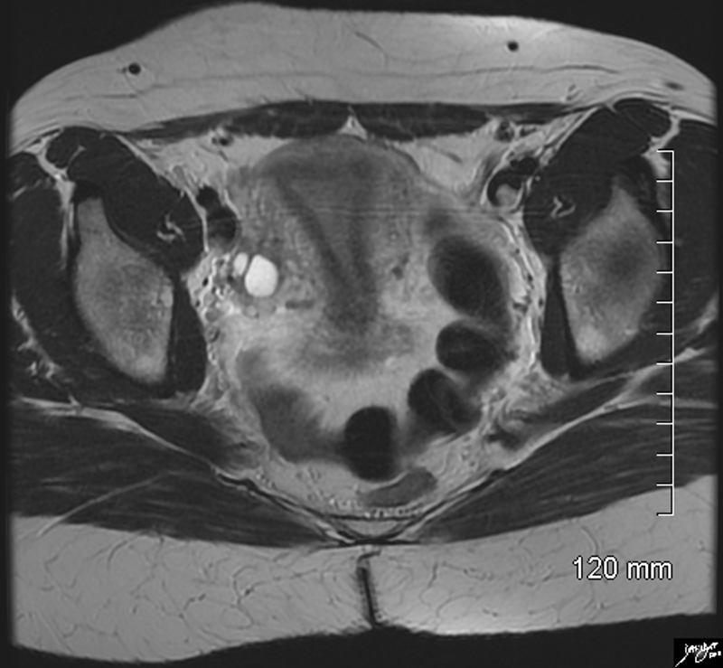
The axial STIR sequence from an MRI study shows the normal appearance of the uterus in a 34 year old female. Because the uterus is anteverted it lies flat so that in this instance the coronal plane of the uterus is demonstrated. The pear shaped form is exemplified together with the 3 parts of the uterus. The inner endometrium of intermediate signal, the junctional zone of low signal consisting of compacted smooth muscle with low water content, and the surrounding myometrium of smooth muscle with a higher water content. Two high signal cuysts are seen in the right ovary Courtesy Ashley Davidoff MD Copyright 2010 All rights reserved 96788.8s |
Hysterosalpingogram – The Irregular Cervical Canal |
|
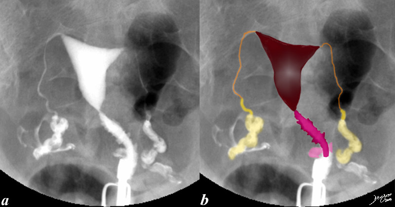
This hysterosalpingogram is from a 26 year old female with normaL endometrial cavity and cervical canal. Note the irregular shape to the cervicalcanal while the endometrial lining is smooth. Note also the free spillage of contrast from the tubes into the peritoneal cavity (white contrast in d, indicating patent tubes. codefallopian tubes and fimbrae Courtesy Ashley Davidoff MD Copyright 2009 all rights reserved 83690c02.8s |

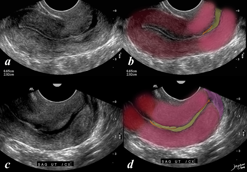
The Flexed Cervix |
|
Normal cervix. Note the flexibility of the cervix and its characteristic serrated appearance of the cavity. Courtesy Ashley Davidoff MD copyright 2009 all rights reserved 30593c.8s |

Normal HSG |
|
Normal cervix. Note the serrated appearance of the cavity uterus cervix normal anatomy HSG hysterosalpingogram Courtesy Ashley Davidoff MD copyright 2010 all rights reserved 30594.8s |
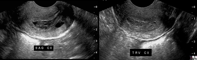
1 Week |
|
26 year old female with normal endometrial cavity and cervical canal filled with echogenic material LMP one week prior Courtesy Ashley Davidoff MD Copyright 2009 all rights reserved 83702.8s |
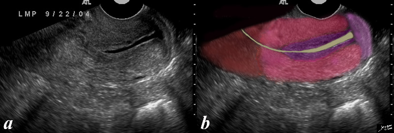
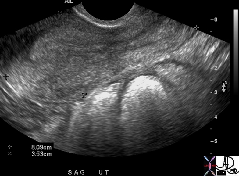
Mid Cycle |
|
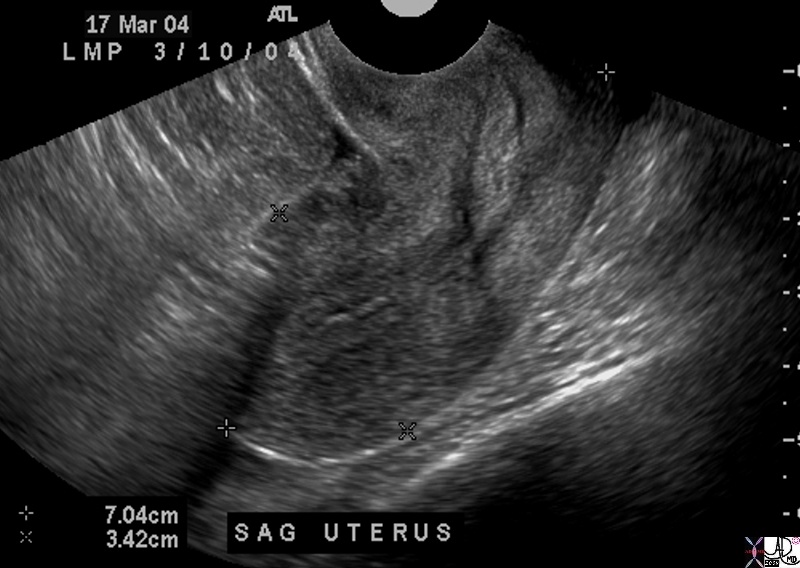
The ultrasound of the normal cervix (salmon pink) is from a 31 year old female whose LMP was 12 days ago, and it shows a fluid filled canal (yellow) surrounded by a thickened mucosa (purple). The lower uterine segment is seen cranially in red and the vagina is seen in pink caudally. Courtesy Ashley Davidoff MD Copyright 2009 all rights reserved 83698c04.8s |
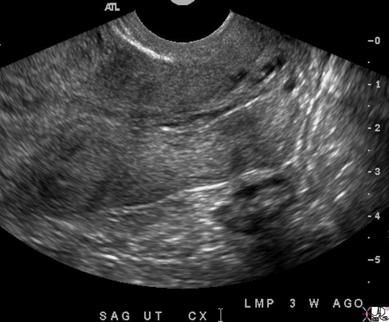
3 Weeks |
|
The ultrasound is from a 42 year old patient who presents witjh pelvic discomfort and an LMP 3 weeks ago. The examination is normal. Nabothian cysts are of incidental note Courtesy Ashley Davidoff MD copyright 2009 all rights reserved 84948.8s |
Mucus – 3 Weeks |
|
25 year old female whose LMP was 10 days ago presents with pelvic pain. The ultrasound shows a normal endometrial cavity and cervical canal filled with simple clear fluid Courtesy Ashley Davidoff MD Copyright 2009 all rights reserved all rights reserved 83689c04.8s |
The Cervix During the Menses |
|
49 year old female presenting with pelvic pain on day 4 of cycle showing shedded endometrial tissue in the cervix. Courtesy Ashley Davidoff MD copyright 2010 all rights reserved 85283c.8s |
Early Pregnancy – Ectopic |
|
The ultrsound is from a young patient presenting with vaginal bleeding for 2 weeks, left lower quadrant pain, and LLQ mass,with a hematocrit of 25, and an HCG 7000. The ultrasound shows no intrauterine pregnancy and a normal endometrial stripe. On a separate image the ectopic pregnancy was noted in the adnexa associated with cystic complex fluid, free fluid and loculated fluid consistent with pelvic hemorrhage. Ectopic pregnancy was diagnosed. The cervical mucosa is thickened and echogenic. Courtesy Ashley Davidoff MD Copyright 2010 46716 |
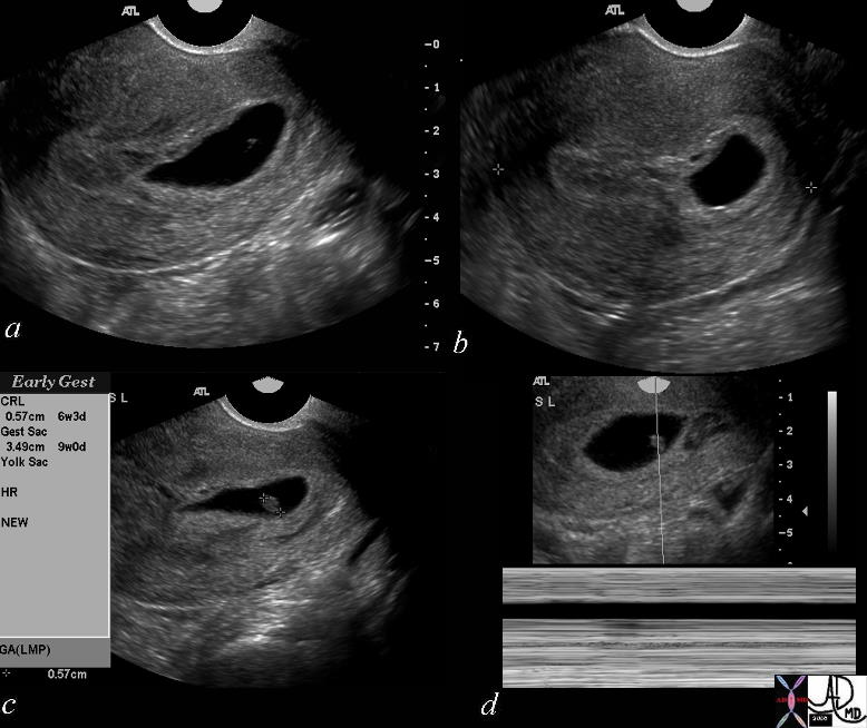
Gestational Sac Bulging into the Cervix Impending Sponataneus Abortion |
|
The history is one of a young female presenting with vaginal bleeding with previous positive pregnancy test. The images show a deformed gestational sac bulging into the cervix. By ultrasound the gestational sac measured 3.49 cms consistent with GA of 9 weeks and CRL measures .49cms consistent with a gestational age of 6weeks and 3 days. No fetal heart was identified. An evolving spontaneous abortion was diagnosed Courtesy Ashley Davidoff MD Copyright 2010 46592c01 |
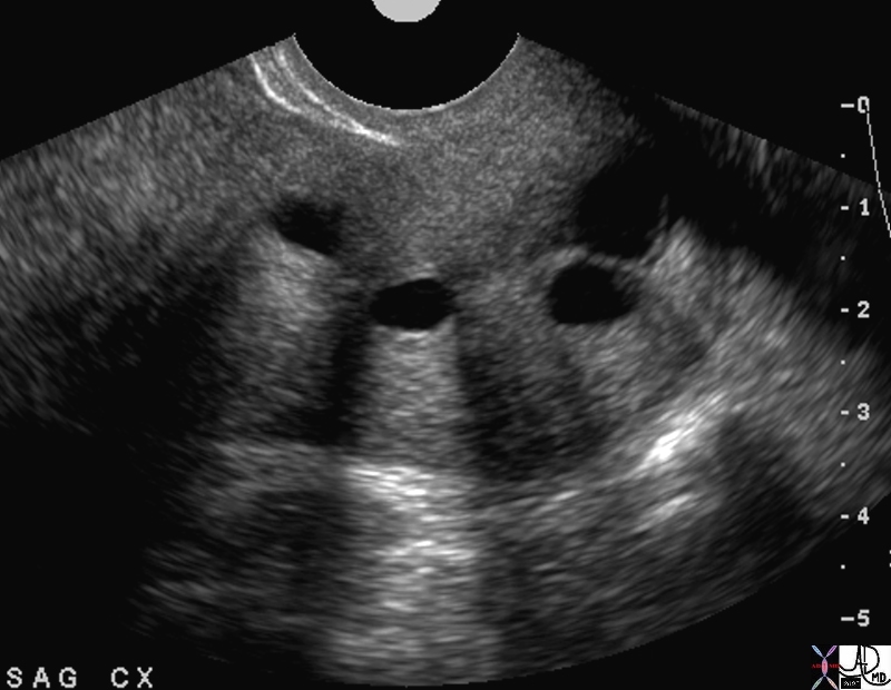
Nabothian Cysts |
| 49463 Courtesy Ashley Davidoff MD |

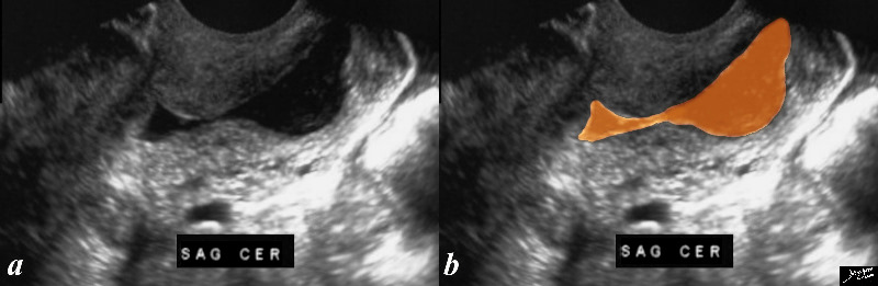
Prolapsing Submucosal Fibroid |
|
This patient presented to the emergency room with severe crampy abdominal pain and a known history of a submucosal fibroid. The MRI shows the fibroid (dark pink) orotruding and expanding the cervix in the sagital view, and in the coronal view it is seen as a hyperemic structure (c) surrounded by lighter pink myometrium. Courtesy Ashley Davidoff MD copyright 2008 16265c01.8s |
Cervical Stenosis and Metrorrhagia |
|
The transvaginal ultrasound is from a 50 year old perimenopausal female with metrorhagia. The uterine cavity and cervical cavity are filled with fluid, and soft tissue elements are identified in the expanded cervical canal. The findings are consistent with cervical stenosis, burt the cause of the metrirhagia is not obvious. The stenosis was relieved and follow up ultrasound showed resolution. No cervical mass was identified. Courtesy Ashley Davidoff MD copyright 2010 all rights reserved 85921c04.8s |
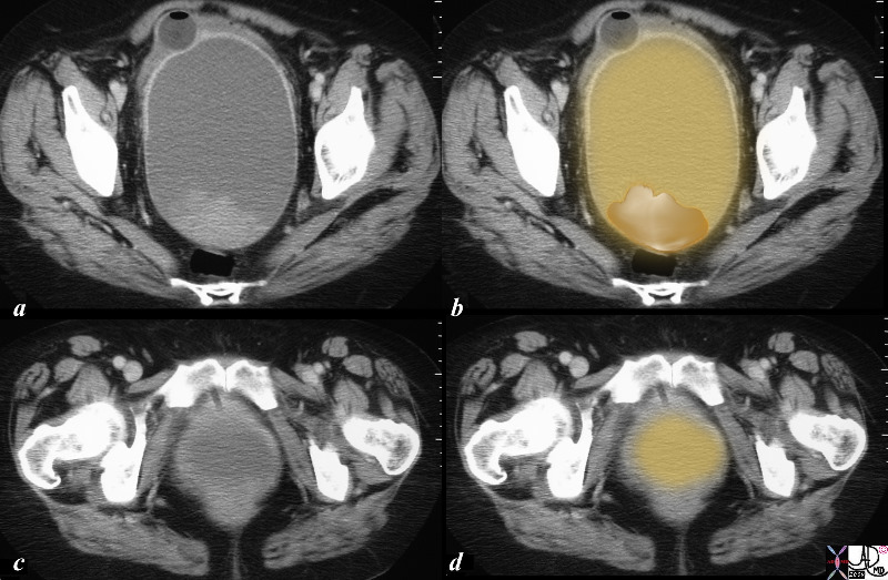
Mucocele of the Cervix |
|
The CTscan is from a 64 year old female who had a cervical mass by clinical examination. The study shows a large cystic mass in the expected location of the cervix. Within the mass there are soft tissue elemets attached to the roof of the mass as well as lying dependantly on the floor of the cyst. A diagnosis of mucocele of the cervix was made following surgery. Courtesy Ashley Davidoff MD copyright 2010 all rights reserved 85931c01.8s |