
Copyright 2010
Niharika Dixit MD Ashley Davidoff MD
Structurally in the non gravid state, the mature uterus is about the size of a woman’s fist, and measures about 8cms X 6cms X 4cms with a volume of about 75-200ccs, and it weighs 100-200gms. It is pear or pyriform shaped muscular organ, and is situated between the bladder anteriorly and the rectum posteriorly. It is a muscular organ with a hollow endometrial cavity. Superiorly the fallopian tubes open on each side into uterine cavity, inferiorly it communicates with vagina through cervix. It consists of a fundus, body (corpus), and neck (cervix).
Histologically the inner endometrial lining consists of a single layer of columnar cells supported by a thin layer of connective tissue, the middle layer is the thickest and is called the myometrium. It consists of smooth muscle and has different layers with various orientation of the fibres. The myometrium proliferates during pregnancy. There is a loose connective tissue layer next which is called the perimetrium and then the outer lining is a cover and is called the peritoneum and is part of a serosal layer. The cyclical changes of the menstrual cycle present a continual change of events controlled by a series of integrated hormonal events. During the follicular phase (proliferative phase) which occurs in the first half of the cycle, and after the shedding of the endometrial lining, there is a rise in estrogen which causes the endometrial lining to start to thicken. In mid cycle after ovulation, luteinizing hormone is released, which heralds in the luteal phase (aka secretory phase). Progesterone now rises and further proliferation of the endometrium occurs. In the absence of pregnancy progesterone levels and estrogen levels fall, and the endometrium sheds.
Common diseases include alterations in the structure which can be congenital or acquired, benign or malignant tumors. Systemic disease especially infections can also affect the uterus and uterine cavity. Diseases include fibroid disease, polyps, adenomyosis, cervical stenosis, and carcinoma. The more common disorders are the functional disorders that relate to cyclical events including menstrual cramps, endometriosis, dysmenorhea, amenorhea, menorhagia. Pain relating to the placement of an intrauterine device is also relatively common.
The developmental uterine anomalies may hinder conception and normal child birth. The changes in position could give rise to chronic pelvic pain. During childbirth there is a risk of injury to urinary bladder as well as anal sphincter as the uterus is anatomically closely related to these structures. The uterus may lose its support with age, repeated pregnancies and post menopause and may give rise to uterovaginal prolapse. Uterine fibroids are most common benign tumors arising from uterine myometrium. Uterine endometrium can stray and become ectopically placed in the myometrium giving rise to adenomyosis and when positioned in the ovaries or pelvis causing a condition called endometriosis .
The diagnosis is dependent initially on clinical evaluation, while the most useful imaging modality is ultrasound. Clinically symptoms include infertility, recurrent abortions, menorrhagia, acute or chronic abdominal pain and urinary complaints. The imaging modalities commonly used include ultrasound, hysterosalpingography, hysteroscopy and diagnostic laparoscopy.
Treatment options are guided by disease process and may include hormonal treatment or minimally invasive or open surgery .
Parts of the Uterus – The Junctional Zone |
|
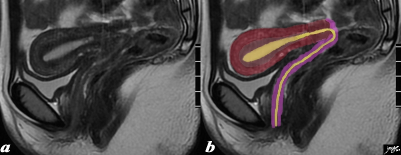
The normal sagittal view of the uterus is a T2 weighted MRI from a 16 year old female with pelvic pain. It demonstrates theta the uterus and more specifically the myometrium is more complex and consists of an outer part (dark red) and an inner more homogeneous part called the junctional zone Since a T2 weighted image is sensitive to water we understand from this image that the outer part since it has greater white signal contains more water, and likely more vascularity. The endometrial canal, cervical canal and vaginal cavity are outlined in yellow and the vaginal wall is pink. Courtesy Ashley Davidoff MD Copyright 2010 All rights reserved 96733c01.8s |
Basic Histology
3 Layers
|
Basic Histological Layers – Body of the Uterus |
|
The wall of the uterus contains 3 basic layers; the mucosa, the muscularis and the serosa as demonstrated in this artistic rendition of the wall of the uterus in the premenstrual phase. Courtesy Ashley Davidoff MD Copyright 2010 All rights reserved 12047e04a03.8s |

Simple Tubular Glands in a Stroma
|
Simple Tubular Gland of the Endometrium |
|
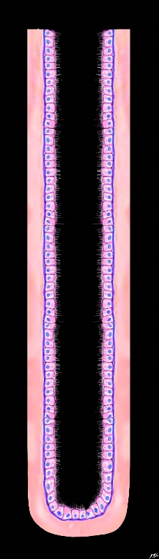
The diagram reveals the histological appearance of the simple tubular gland of the endometrial lining of the uterus The ciliated columnar cells rest on a basement membrane (deep blue) which in turn are embedded in a stroma (pink) Courtesy Ashley Davidoff MD Copyright 2010 All rights reserved 32347f02.8s |
Imaging the Endometrium – Ultrasound
The Complex Endometrium Triple Stripe – Trilaminar Appearance – Preovulation |
|
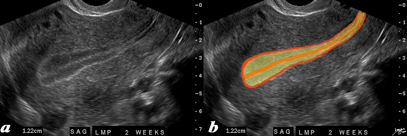
The normal sagittal view of the uterus is a transvaginal ultrasound, 2 weeks after menstruation, and just prior to ovulation. It demonstrates that the uterus and more specifically the endometrium is more complex than just a simple columnar epithelium. This is an example of the trilaminar appearance of the endometrium and is characteristic of the appearance of the endometrium in the preovulatory phase. This pahse is also called the follicular phase, or proliferative phase. It is during this time that estrogen is the dominant hormonal influence. The appearance is also known as a “triple stripe”, and conversely, its presence is seen before progesterone is produced. If present with a 9mm+ diameter, it reflects an ideal potential lining for fertilization.. In this case it measured 1.2cms. Courtesy Ashley Davidoff MD Copyright 2010 All rights reserved 83835c02.8s |
The Overipe Endometrium – Premenstrual |
|
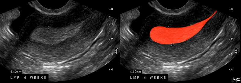
The normal sagittal view of the uterus is a transvaginal ultrasound, about 4 weeks after menstruation, and just prior to the next menstrauation when the endometrium is overripe. It demonstrates that the uterus and more specifically the endometrium is more complex than just a simple columnar epithelium. This is an example of the hyperechoic, homogeneous, thick endometrium characteristic of the secretory phase. It is during this time that progesterone is the dominant hormonal influence and estrogen influence is minimal. The endometrium in this case measures 1.2cms. Courtesy Ashley Davidoff MD Copyright 2010 All rights reserved 46318c01.8s |
Life Depends on the Uterus
A Reason For Living |
|
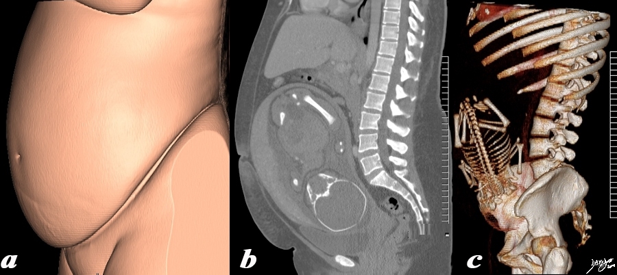
The 3 sagittally reconstructed images of the gravid uterus are from a normal 31 year woman carrying a 32 week gestation. They depict the outside appearance of the patient (a), 2 D sagittal view (b) and 3D sagittal view(c). Pregnancy is the main reason for living for the uterus and the opportunity to use it for this purpose is only a brief time in the long life of an adult female – but the species depends on this brief sojourn. Courtesy Ashley Davidoff MD Copyright 2010 All rights reserved 96332c.91s |