
Copyright 2009
Ashley Davidoff MD
Definition
The endometrial stripe is a dynamic structure subject to the fluxesof the hormones in the days preceding ovulation and the days of expectant implanation in the days following ovulation.
|
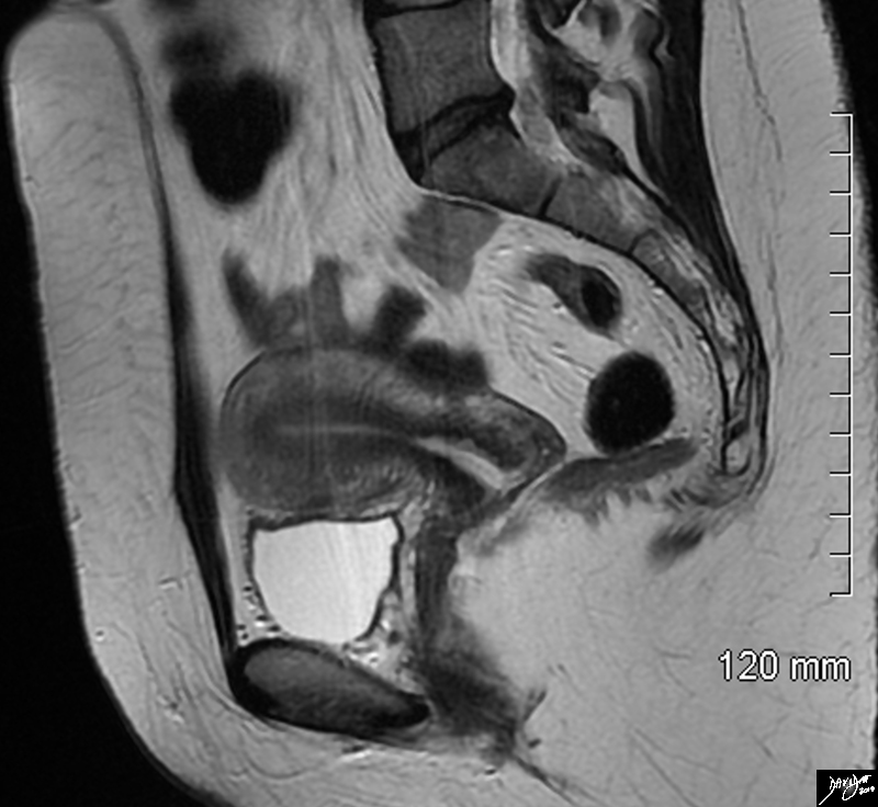
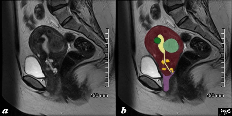
The Uterus in the Sagittal Plane |
|
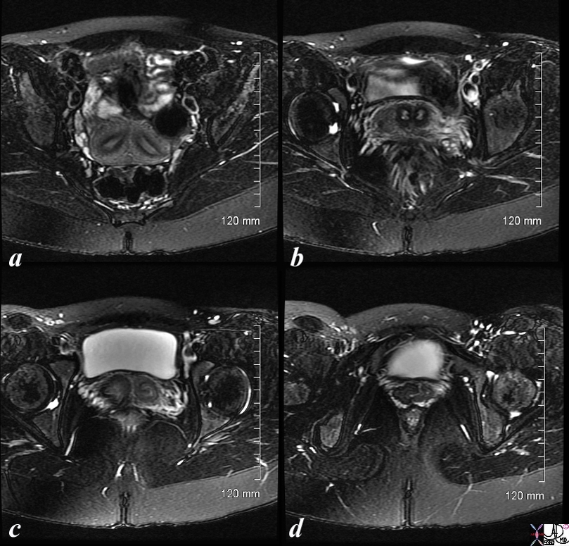
The sagittal STIR sequence from an MRI study shows the normal appearance of the uterus in a 34 year old female. The pear shaped form is exemplified together with the 3 parts of the uterus. The inner endometrium of intermediate signal, the junctional zone of low signal consisting of compacted smooth muscle with low water content, and the surrounding myometrium of smooth muscle with a higher water content. Courtesy Ashley Davidoff MD Copyright 2010 All rights reserved 96785.8s |
|
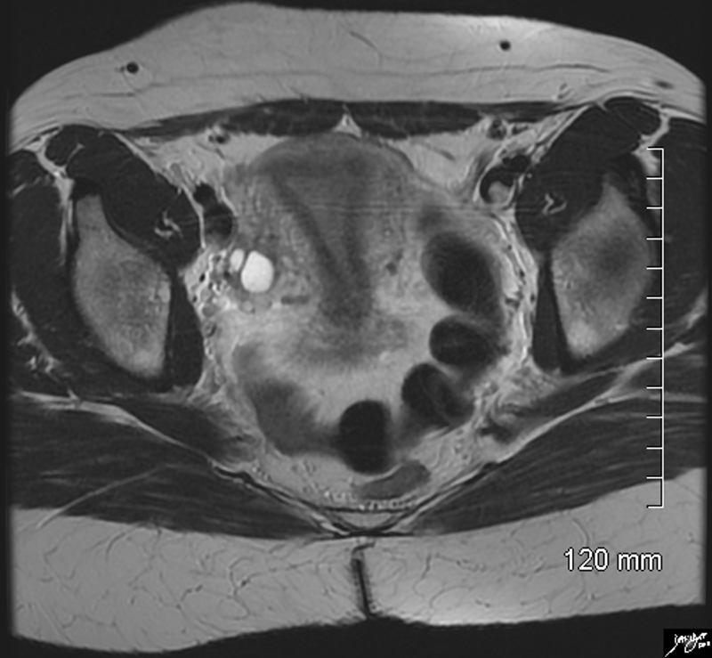
Axial Plane Coronal Projection |
|
The axial STIR sequence from an MRI study shows the normal appearance of the uterus in a 34 year old female. Because the uterus is anteverted it lies flat so that in this instance the coronal plane of the uterus is demonstrated. The pear shaped form is exemplified together with the 3 parts of the uterus. The inner endometrium of intermediate signal, the junctional zone of low signal consisting of compacted smooth muscle with low water content, and the surrounding myometrium of smooth muscle with a higher water content. Two high signal cuysts are seen in the right ovary Courtesy Ashley Davidoff MD Copyright 2010 All rights reserved 96788.8s |
In the follicular phase, during the days when estrogen influence dominates the
In the preovulatory phase a trilaminar endonetrial stripe is present characteristic of the proliferative phase
The luteal phase is dominated by progesterone influence changing the endometrium into a secretory pattern characterized by ultrasound as a increasingly echogenic and lss of the trilaminar pattern
1 week
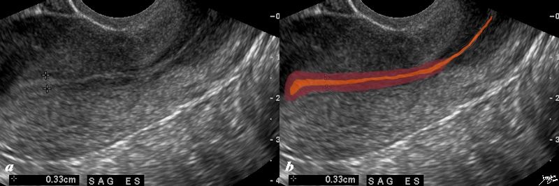
Endometrium In Early Proliferative Phase |
|
The normal sagittal view of the uterus is a transvaginal ultrasound, in the first week after menstruation after menstruation, and just prior to the next menstruation after the endometrium has been shed. It demonstrates that the endometrium becomes a single echogenic line consisting of opposing walls (orange) and is surrounded by a subendometrial halo of the junctional zone (tan). This layer is more compacted, and relatively hypovascular. This image is typical of the early proliferative phase. It is during this time that estrogen starts to rise and progesterone has fallen. The endometrium in this case measures about 3mms. Courtesy Ashley Davidoff MD Copyright 2010 All rights reserved 84698c02b.8s |
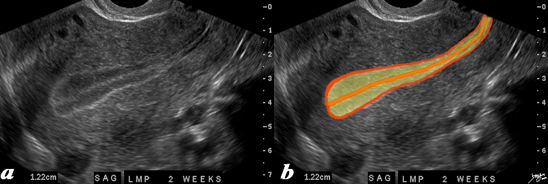
2 Weeks
|
Endometrium in LAte Proliferative Phase Triple Stripe – Trilaminar Appearance – Preovulation |
|
The normal sagittal view of the uterus is a transvaginal ultrasound, 2 weeks after menstruation, and just prior to ovulation. It demonstrates that the uterus and more specifically the endometrium is more complex than just a simple columnar epithelium. This is an example of the trilaminar appearance of the endometrium and is characteristic of the appearance of the endometrium in the preovulatory phase. This pahse is also called the follicular phase, or proliferative phase. It is during this time that estrogen is the dominant hormonal influence. The appearance is also known as a “triple stripe”, and conversely, its presence is seen before progesterone is produced. If present with a 9mm+ diameter, it reflects an ideal potential lining for fertilization.. In this case it measured 1.2cms. Courtesy Ashley Davidoff MD Copyright 2010 All rights reserved 83835c02.8s |
3 Weeks
|
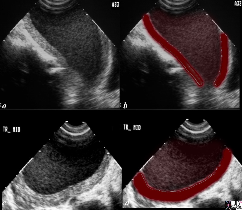
Endometrium in Mid Secretory Phase LMP about 3 Weeks Prior |
|
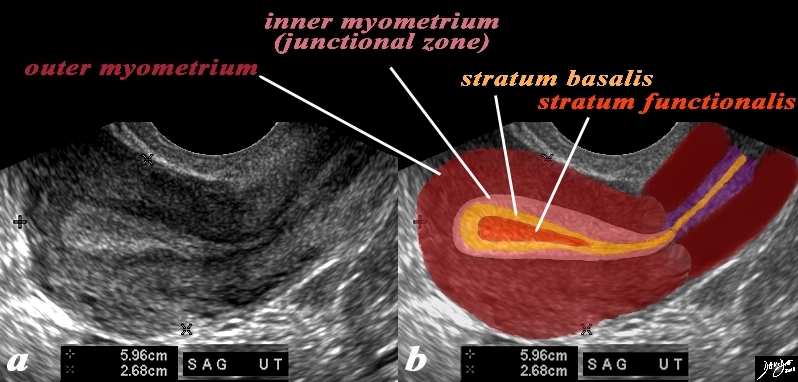
In this 26 year premenstrual female a transvaginal ultrasound in the sagittal plane reveals a normal view of the uterus with characteristic premenstrual appearance. (a) The stripe is almost homogeneously echogenic and thick but also shows a hypoechoic halo of the junctional zone or inner myometrium. (salmon) The homogeneous stripe is made up from two histological layers (barely distinguished by this ultrasound)– the inner stratum functionalis (deep orange) that will shed once the spiral arteries vasoconstrict, and the outer stratum basalis (deep yellow) that will not shed, and will be the basis for regenerating the endometrium in the next cycle. The next layer as stated above is the compact myometrium – the junctional zone (aka inner myometrium) , and is followed by the thicker outer myometrium (maroon). (b) Courtesy Ashley Davidoff MD Copyright 2010 All rights reserved 84538c06.83s |
4 Weeks
Premenstrual |
|
The normal sagittal view of the uterus is a transvaginal ultrasound, about 4 weeks after menstruation, and just prior to the next menstrauation when the endometrium is overripe. It demonstrates that the uterus and more specifically the endometrium is more complex than just a simple columnar epithelium. This is an example of the hyperechoic, homogeneous, thick endometrium characteristic of the secretory phase. It is during this time that progesterone is the dominant hormonal influence and estrogen influence is minimal. The endometrium in this case measures 1.2cms. Courtesy Ashley Davidoff MD Copyright 2010 All rights reserved 46318c01.8s |
High Ppower Changes at Menstruation Shedding the Functional Layer |
|
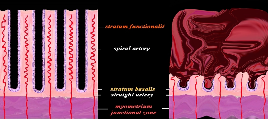
The diagram reflects the premenstrual endometrium (left) and the post menstrual endometrium right, revealing the necrosis of the functional layer with sloughing and hemorrhage. The basal layer with the staright arteries and a small portion of the spiral artery remains intact. The hemorrhage is controlled by spasm of the arteries and contraction of the myometrium. Courtesy Ashley Davidoff MD Copyright 2010 All rights reserved 32347f08cL.92s |
Endometrium During Pregnancy
|
Junctional Zone – Early Pregnancy |
|
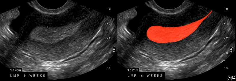
In this 16 year old patient her LMP was about 4 weeks ago and she had a positive pregnancy test. The transvaginal ultrasound in the sagittal plane reveals a normal view of the uterus with characteristic early pregnancy appearance characterized by the gestational sac (gs) embedded in the stratum functionalis (deep orange). The stratum basalis is seen as a slightly more echogenic layer around the functional layer (deep yellow). The stripe is thick measuring about The next layer is a barely seen junctional layer (salmon pink) best seen on the anterior subendometrial layer just beyond the basalis The next layer is the thicker outer myometrium (maroon) that contains dilated vessels. Courtesy Ashley Davidoff MD Copyright 2010 All rights reserved 84485c02.8ls |
Post Menopausal
|
Post Menopausal uterus Junctional Zone Prominent |
|
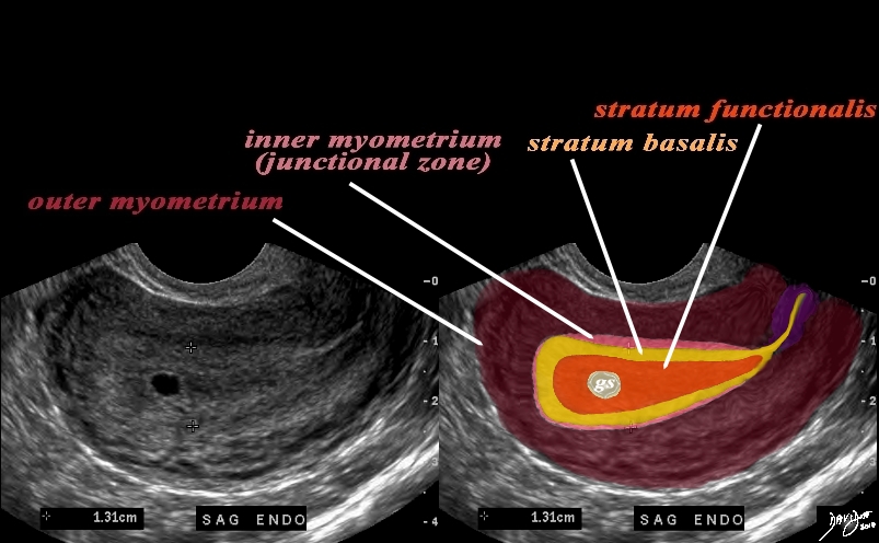
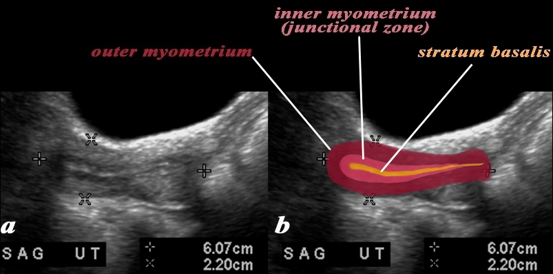
In this 60 year old female with normal atrophied post menopausal uterus the transabdominal ultrasound in the sagittal plane reveals a normal view of the uterus with characteristic postmenopausal appearance. The stripe is thin measuring about 2mm homogeneously echogenic but also shows a hypoechoic halo of the junctional zone or inner myometrium. (salmon) The echogenic stripe is made up from a single histological layer the stratum basalis (deep yellow) The next layer as stated above is the compact myometrium – the junctional zone (aka inner myometrium) , and is followed by the thicker outer myometrium (maroon). Courtesy Ashley Davidoff MD Copyright 2010 All rights reserved c uterus USscan Courtesy Ashley Davidoff MD Copyright 2009 all rights reserved 83573c01.81L |
|
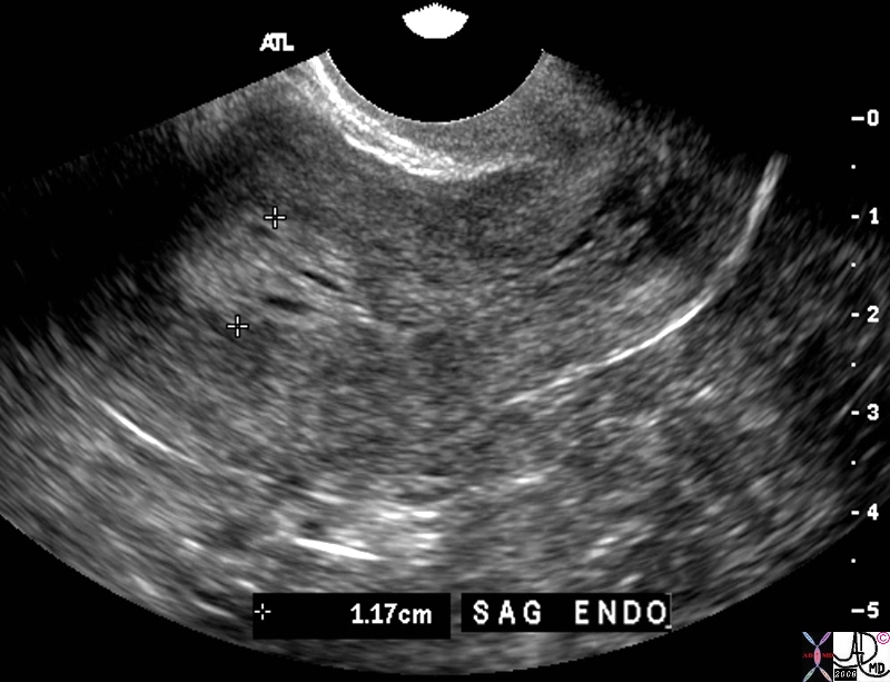
Submucosal Fibroid |
| The USscan (sonohysterogram) scan is from a 41year old female, who presents with dysfunctional uterine bleeding. The study reveals a 4mms submucosal nodule shown at pathology to be a submucosal fibroid.
uterus fibroid leiomyoma submucosal USscan sonohysterogram Courtesy Ashley Davidoff MD copyright 2009 all rights reserved 84299c041.8 |
|
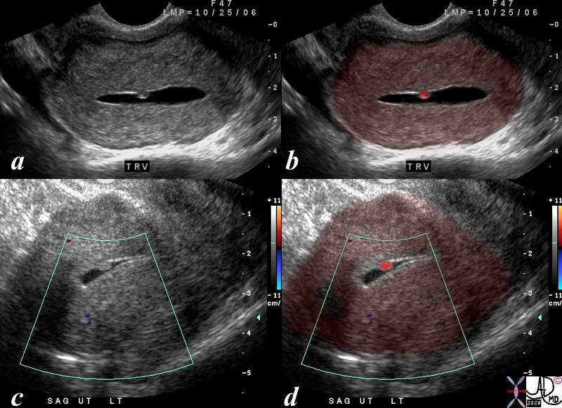
Hyperplastic Polyp |
| 84299c05.8s The USscan (hysterosonogram is from a 47year old female, with history metrorhagia dysfunctional uterine bleeding. The studies reveal a mass in the uterus within the endometrial cavity The polypwas shown to be a benign hyperplastic polyp. uterus endometrium mass polyp hyperplastic USscan hysterosonogram Courtesy Ashley Davidoff MD copyright 2009 all rights reserved |
Filling Defect |
|
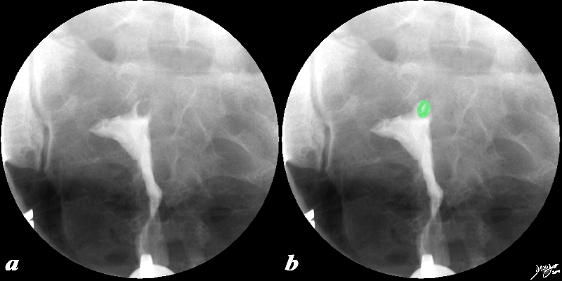
This 38 year old female presents with a history of infertility. The hysterosalpingogram shows non filling of both Fallopian tubes and a filling defect (green) in the region of the patient’s left cornu that likely is the cause of the obstructed left tube and in part the cause of the infertility. The lesion is most likely a submucosal fibroid. Courtesy Ashley Davidoff MD Copyright 2010 All rights reserved 96897c02.8s |
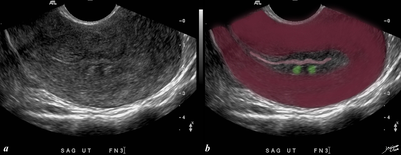
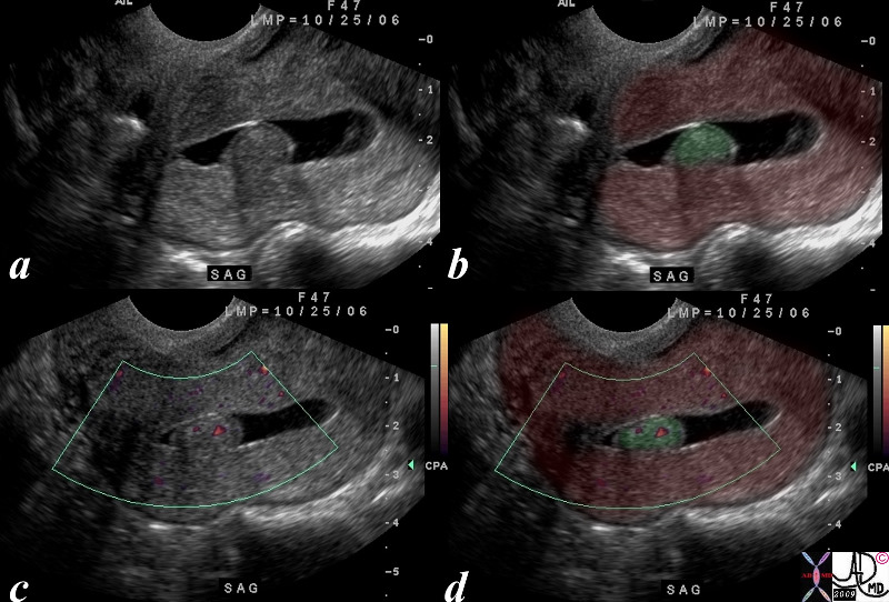
Two Lesions in the Junctional Zone Impinging on the Endometrium Subucosal Fibroid or Adenomyosis |
| This patient presents with menorrhagia. Two echogenic nodules (overlaid in green) are seen in the subendometrial layer, (junctional zone). They are in close proximity and do appear to have appositional relationships with the endometrial stripe. They appear to and distort the endometrial lining. These findings could account for the patient’s menorrhagia. Note that the uterus is retroverted Included in the differential diagnosis are submucosal fibroids, and dystrophic changes in prior foci of adenomyosis. An MRI would be helpful in further characterizing these lesions in the subendometrial layer
copyright 2009 all rights resrved Courtesy Ashley Davidoff MD 85641bc01.8s |
Hyperplasia
Thick Heterogeneous Endometrium |
|
The transvaginal ultrasound is from a 60 year old female who presents with spotting Ultrasound reveals a heterogeneous endometrial stripe consistent with endometrial hyperplasia though endometrial carcinoma is a possibility. Malignant neoplasia is a a les likely possibility Courtesy Ashley DAvidoff MD copyright 2010 83301.81s |
Endometrial Cancer
|
Endometrial Carcinoma and Uterine Obstruction |
|
The ultrasound is from a 70 year old post menopausal female who presents with an enlarged uterus. The endometrial stripe is enlarged and is filled with fluid and an enhancing soft tissue mass consistent with an endometrial carcinoma. Note blood flow as depicted by Doppler exam (c) characterizing the soft tissue as tumor rather than a clot. Courtesy Ashley Davidoff MD copyright 2009 all rights reserved 86206c.8s |
|
Focal Endometrial Thickening |
|
The coroanal CTreformatted CTscan images are from a 46 year old female with endometrial carcinoma. The images show an focal area of soft tissue thickening (green), on the superior surface (b) and rightward (green d) of the endometrial cavity (orange) . 83494c01.8s Courtesy Ashley Davidoff Copyright 2010 all rights reserved |

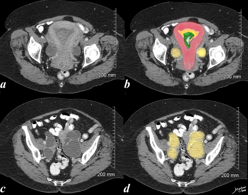
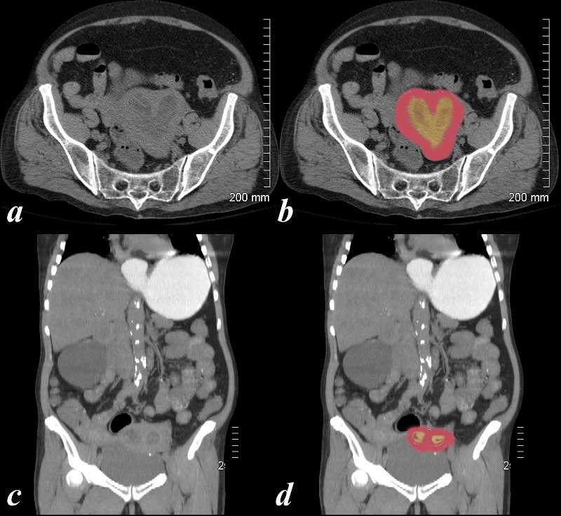
Endometrial Carcinoma with Obstruction of the Uterus and Fallopian Tubes |
| 70 year old female with pelvic discomfort. The CT shows an endometrium filled with complex soft tissue (green), fluid and or blood (orange) and the Fallopian tubes which are distended with fluid (yellow) caused by the obstructing carcinoma.
Courtesy Ashley DAvidoff MD 48414c02.8s |
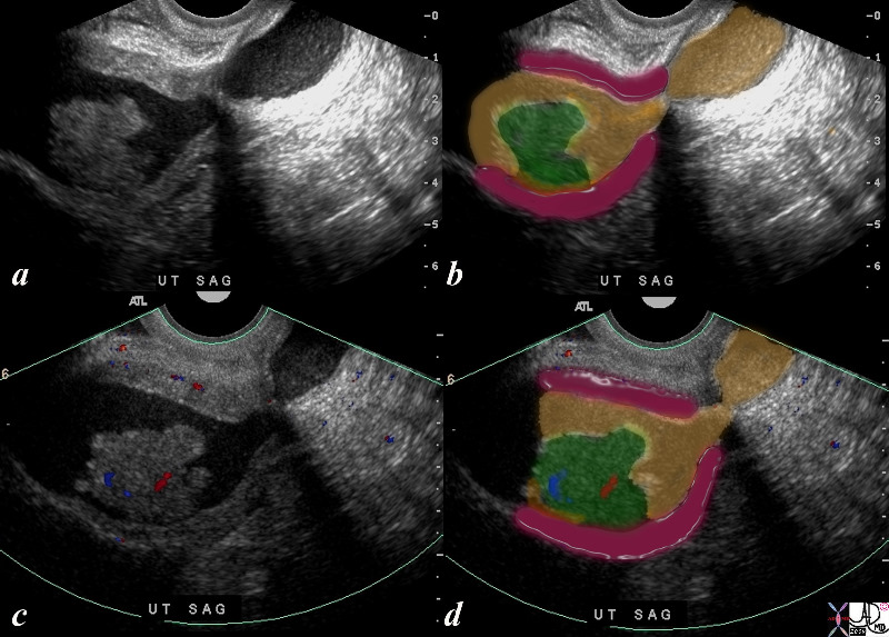
Fibroid Impingement on the Endometrium |
|
In this sagittal view of a T2 weighted MRI of the uterus of a 42 year old female with multiple fibroids two of which impinge on the endometrial cavity (the larger (light green) from posterior and the smaller (dark green) from anterior resulting in a sigmoid shaped cavity (yellow). In addition incidentally noted Nabothian cysts (orange) are seen within the cervix. Courtesy Ashley Davidoff MD Copyright 2010 All rights reserved 96531c02.8s |
|
Hemorrhage into the Endometrial Cavity |
| The ultrasound is from a 33 year old female who had a cervical biopsy and then showed complex fluid in a distended endometrial cavity. the findings are consistent with hematocolpos, but in the appropriate clinical setting could represent pyocolpos
Courtesy Ashley Davidoff MD copyright 2009 all rights reserved 85940c02.8s |
|
Deformity of the Endometrial CAvity Caused by an Intramural Fibroid |
|
In this sagittal view of the T1 weighted contrast enhanced study of the the uterus of a 46 year old female. The uterus roughly retains its pear shaped structure but the shape of the endometrial cavity is distinctly abnormal as if there is something pushing on it from above. This patient has a diffusely enhancing isointense leiomyoma that impinges on the endometrial cavity. Courtesy Ashley Davidoff MD Copyright 2010 All rights reserved 96577.8s |
|
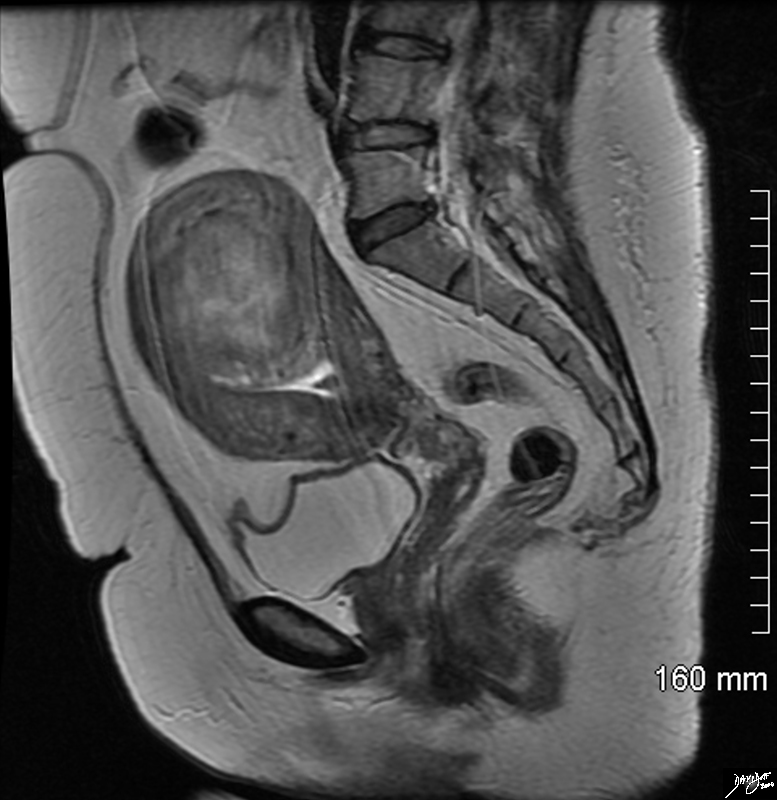
Large Intramural Fibroid Deforming the Stripe T2 Weighted Image |
|
In this sagittal view of the T2 weighted MRI of the uterus of a 46 year old female an almost 6cms slightly hyperintense fibroid is seen pushing on the endometrial cavity from above. This patient has a large fibroid aka leiomyoma that impinges on the endometrial cavity. Courtesy Ashley Davidoff MD Copyright 2010 All rights reserved 96570.8s |
|
Carcinoma of the Cervix in a Bicornuate Uterus Complicated by Obstruction |
| 74 year old female with bicornuate uterus and dilated endometrial cavities Diagnosis is carcinoma of the cervix with obstruction . The myometrium is overlaid in dark pink, and the endometrial cavity is a heterogeneous orange consisting of both fluid and soft tissue elements.
Courtesy Ashley Davidoff MD copyright 2009 all rights reserved 83438c01.8s |
Congenital Anomalies
|
Two Hemiuteri, Two Cervices and One Vagina MRI T2 Weighted with FAt Saturation |
|
The MRI is from a 24F year old female with uterus didelphys The T2 weighted study in axial projection reveals 2 uterine corpuses (a) 2 cervices (b), vaginal septum in the upper 1/3 (c) but single distal vagina (d) Image Courtesy Ashley Davidoff MD Copyright 2010 83730c01L.8s |
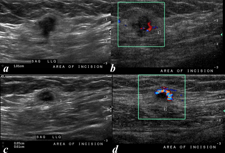
Ectopic Endometrium |
| 35 year old female who is post C section shows a nodule within the scar of the cesarian section most consistent with an endometrial implant. The ultrasound shows two nodules, each with doppler flow, characteristic in appearance and by location of ectopic endometrial tissue.
Courtesy Ashley Davidoff MD Copyright 2009 all rights reserved 83624c01.8 |
References
Nalaboff, Kenneth M. MD, Pellerito, John S. MD and Ben-Levi, Eran MD Imaging the Endometrium: Disease and Normal Variants RadioGraphics, 21, 1409-1424. November 2001
E. R. Sokol , H. Casele and E. I. Haney Ultrasound examination of the postpartum uterus: what is normal?
2004, Vol. 15, No. 2 , Pages 95-99