
Nerve Supply
Introduction
The innervation of the uterus is entirely autonomic arising from the inferior hypogastric plexus (sympathetic) and the pelvic splanchnic nerves (parasympathetic from S2,3,4). The afferent fibres travel with the sympathetic efferents to T10-12 and L1 spinal cord segments.
In the sympathetic chain there is an intermediary step between the viscera and spinal cord – a “halfway house” if you will, that runs alongside the spinal cord, and it is called the sympathetic chain. There is therefore a neuron that carries the signal from the spinal cord to the sympathetic chain called the preganglionic neuron, and one that carries the signal between the ganglion of th chain to the organ called the post ganglionic neuron In general T1 goes to head, T2- T6 the thorax and T7 -T11 the abdomen. T12 , L1 and L2 go to the legs. This is an approximation and there is a large cross over. The innervation depends on the embryological origin. Since the hear originated in the neck of the fetus the innervation is from the neck. Similarly the foregut organs arose from the chest endomesoderm and so innervation is from the lower thoracic region
Most the afferent fibres ascend through the inferior hypogastric plexus and enter the spinal cord via T10-12 and L1 spinal nerves.
The nerve supply to the uterus is derived from the hypogastric and ovarian plexuses, and from the third and fourth sacral nerves. The sympathetic drive to the uterus helps in relaxation of uterine muscle while parasympathetic supply.
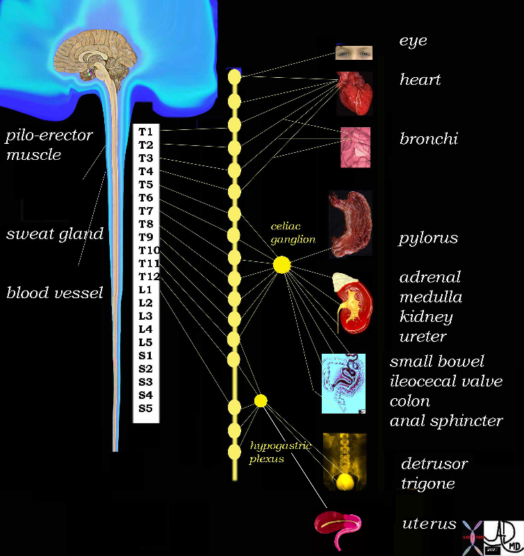
Sympathetic System |
|
The diagram reveals the sympathetic nervous system of the body and in this instance attention is drawn to the contribution of the hypogastric plexus, and specifically from a component called the inferior hypogastric plexus which in turn gets contributions from the anterior and intermediate part known as the uterovaginal plexus. The plexus lies in the broad ligament on each side of the cervix. Courtesy Ashley Davidoff MD Copyright 2010 All rights reserved v |
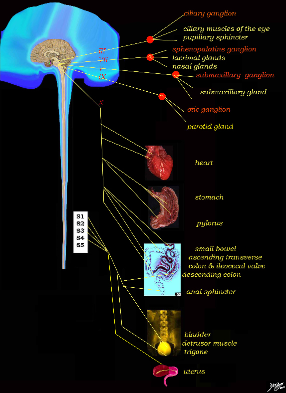
Parasympathetic System |
|
The diagram reveals the parasympathetic nervous system of the body and in this instance attention is drawn to the contribution of the S2,3 and 4, called the pelvic splanchnic plexus Courtesy Ashley Davidoff MD Copyright 2010 All rights reserved 14798d05e01.8s |
References
O’Rahilly, Müller, Carpenter & Swenson Dartmouth Anatomy Text