
The Common Vein Copyright 2010
Introduction
|
Normal Gray Scale and Power Doppler |
| 46314c01 uterus endometrium blood flow myometrium perfusion normal anatomy USscan Davidoff MD |
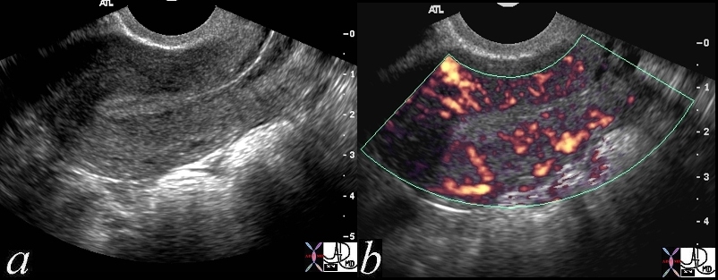
Junctional Zone and Outer Myometrium |
|
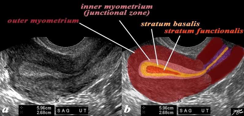
In this 26 year premenstrual female a transvaginal ultrasound in the sagittal plane reveals a normal view of the uterus with characteristic premenstrual appearance. (a) The stripe is almost homogeneously echogenic and thick but also shows a hypoechoic halo of the junctional zone or inner myometrium. (salmon) The homogeneous stripe is made up from two histological layers (barely distinguished by this ultrasound)– the inner stratum functionalis (deep orange) that will shed once the spiral arteries vasoconstrict, and the outer stratum basalis (deep yellow) that will not shed, and will be the basis for regenerating the endometrium in the next cycle. The next layer as stated above is the compact myometrium – the junctional zone (aka inner myometrium) , and is followed by the thicker outer myometrium (maroon). (b) Courtesy Ashley Davidoff MD Copyright 2010 All rights reserved 84538c06.83s |
Transverse Section Ultrasound |
|
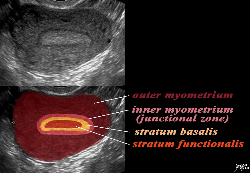
In this 26 year premenstrual female a transvaginal ultrasound reveals a normal transverse view of the uterus with characteristic premenstrual appearance. The stripe is homogeneously echogenic and thick but also shows a hypoechoic halo of the junctional zone or inner myometrium. (salmon) The homogeneous stripe is made up from two histological layers (not distinguished by this ultrasound)– the inner stratum functionalis (deep orange) that will shed once the spiral arteries vasoconstrict, and the outer stratum basalis (deep yellow) that will not shed, and will be the basis for regenerating the endometrium in the next cycle. The next layer as stated above is the compact myometrium – the junctional zone (aka inner myometrium) , and is followed by the thicker outer myometrium (maroon). Courtesy Ashley Davidoff MD Copyright 2010 All rights reserved 84540cc06.8s |
CT in Sagittal Section |
|
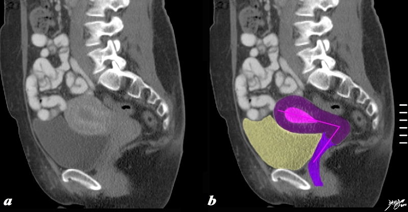
The sagittaly reconstructed CT shows an anteverted uterus buoyed and cushioned by partly filled bladder (yellow) In this sagittal view of the uterus an ‘L” shaped structure of the uterus vagina, and the internal cavity is diagrammed in by the pink vectors of the tract. Courtesy Ashley Davidoff MD copyright 2010 all rights reserved 60385c04b.8s |
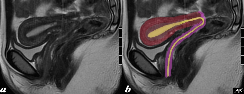
STIR Weighted Image in Sagittal Projection |
|
The sagittal STIR sequence from an MRI study shows the normal appearance of the uterus in a 34 year old female. The pear shaped form is exemplified together with the 3 parts of the uterus. The inner endometrium of intermediate signal, the junctional zone of low signal consisting of compacted smooth muscle with low water content, and the surrounding myometrium of smooth muscle with a higher water content. Courtesy Ashley Davidoff MD Copyright 2010 All rights reserved 96785.8s |