
The Common Vein Copyright 2010
Definition
Postmenopausal uterine bleeding is bleeding that occurs in a woman whose menstrual periods have ceased for 12 months and has varying causes.
The most common cause is atrophy of the vaginal mucosa or endometrium. However, endometrial cancer causes 10-15% of cases of postmenopausal bleeding.
Structural and functional changes vary depending on cause.
However, the result is always uterine bleeding after menopause, which can resemble menstrual flow, giving women their common clinical presentation.
Diagnosis is based on clinical history, and the evaluation should include endometrial biopsy to rule out endometrial cancer.
Treatment depends on the cause found at evaluation.
Atrophy
Atrophic Endometrium |
|
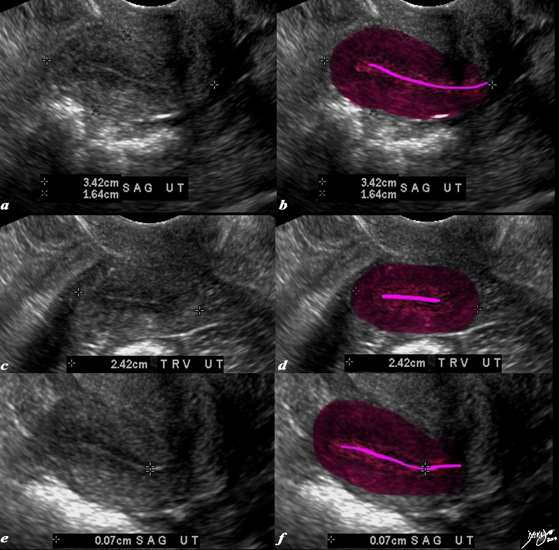
The transvaginal ultrasound is from a 62 year old post menopausal patient who presents with pelvic discomfort. The ultrasound shows a small atrophied uterus. The myometrium (dark red) junctional zone (light red) and endometrial cavity (pink) are outlined. In the sagittal plane the uterus measures 3.4cms in cranoicaudal dimension by 2.4cms in anteroposterior dimension (a,b) In the axial dimension (c,d) the uterus measures 2.4cms. The endometrial stripe is measured in the sagittal plane and measures .7mms. Courtesy Ashley Davidoff MD Copyright 2010 All rights reserved 84711c01.8s |
Endometrial Hyperplasia
|
Thick Heterogeneous Endometrium |
|
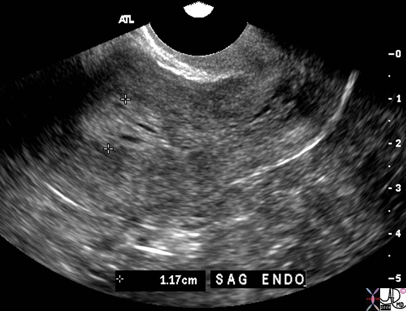
The transvaginal ultrasound is from a 60 year old female who presents with spotting Ultrasound reveals a heterogeneous endometrial stripe consistent with endometrial hyperplasia though endometrial carcinoma is a possibility. Malignant neoplasia is a a les likely possibility Courtesy Ashley DAvidoff MD copyright 2010 83301.81s |
Endometrial Carcinoma
|
Endometrial Carcinoma and Uterine Obstruction |
|
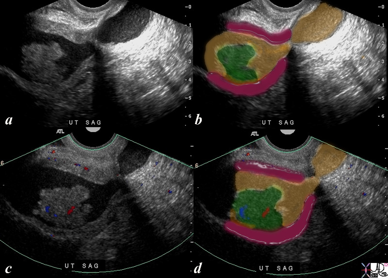
The ultrasound is from a 70 year old post menopausal female who presents with an enlarged uterus. The endometrial stripe is enlarged and is filled with fluid and an enhancing soft tissue mass consistent with an endometrial carcinoma. Note blood flow as depicted by Doppler exam (c) characterizing the soft tissue as tumor rather than a clot. Courtesy Ashley Davidoff MD copyright 2009 all rights reserved 86206c.8s |
|
Focal Endometrial Thickening |
|
The coroanal CTreformatted CTscan images are from a 46 year old female with endometrial carcinoma. The images show an focal area of soft tissue thickening (green), on the superior surface (b) and rightward (green d) of the endometrial cavity (orange) . 83494c01.8s Courtesy Ashley Davidoff Copyright 2010 all rights reserved |
