
Copyright 2009
Introduction
Indications
Uterine masses
Pelvic pain or vaginal bleeding
Congenital abnormalities
Technique
Cor T2 SSFSE add kidneys
T1 SE or GRE +/- Fat suppression
T2 axial sagittal coronal
Uterine axis
Pre and post gad T1 SPGR FS
Results
Uniform T1 poor tissue contrast
Best seen on T2
endometrium bright
Junctional zone is dark
Myometrium intermediate
T1 with Gadolinium
|
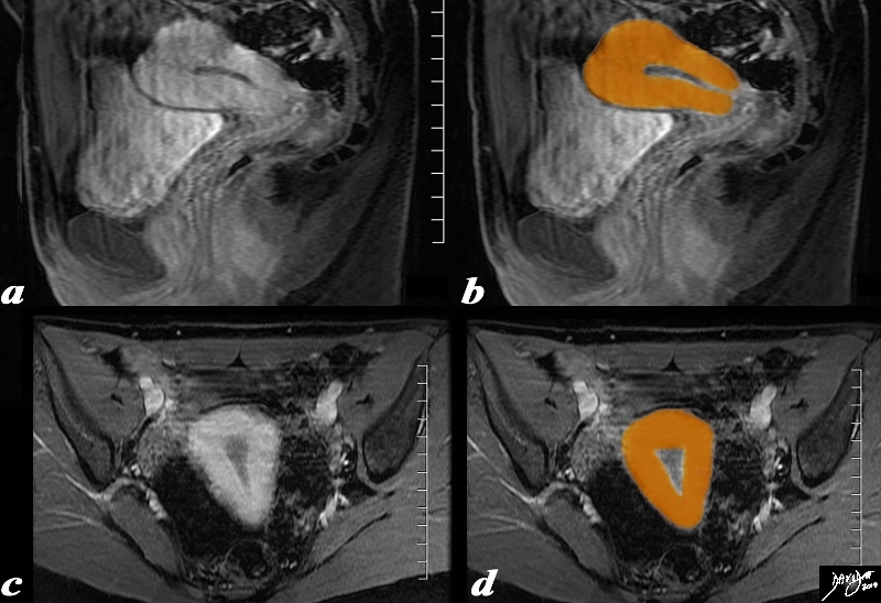
Pear Shape in both the Sagital and Coronal Planes |
|
The uterus is pear shaped as depicted in this overlay sagital (a,b) and coronal (c,d) T 1weighted enhanced MRI study Courtesy Ashley Davidoff MD Copyright 2010 96378c02.81s |
STIR
|
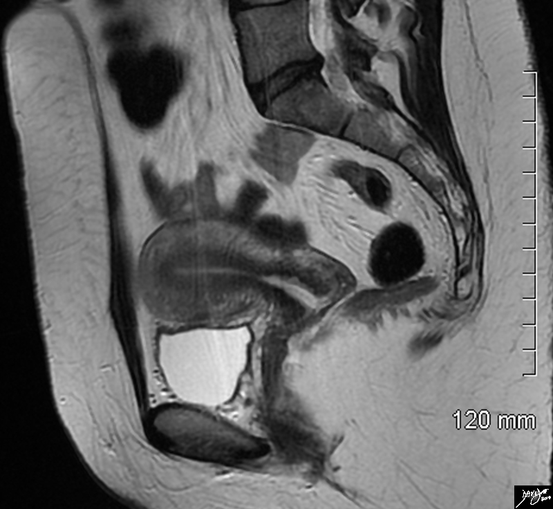
The Uterus in the Sagittal Plane |
|
The sagittal STIR sequence from an MRI study shows the normal appearance of the uterus in a 34 year old female. The pear shaped form is exemplified together with the 3 parts of the uterus. The inner endometrium of intermediate signal, the junctional zone of low signal consisting of compacted smooth muscle with low water content, and the surrounding myometrium of smooth muscle with a higher water content. Courtesy Ashley Davidoff MD Copyright 2010 All rights reserved 96785.8s |
|
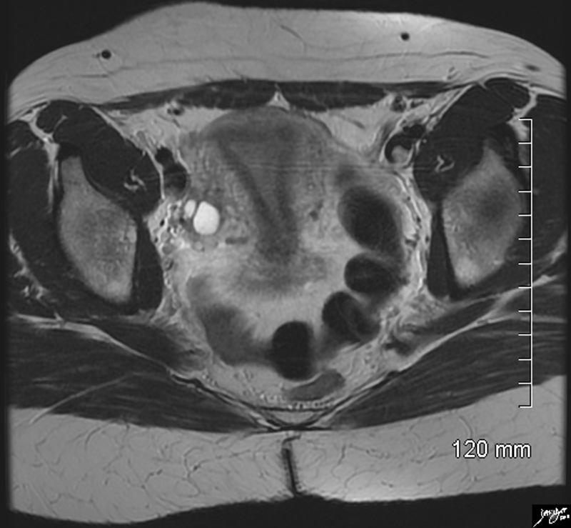
Axial Plane Coronal Projection |
|
The axial STIR sequence from an MRI study shows the normal appearance of the uterus in a 34 year old female. Because the uterus is anteverted it lies flat so that in this instance the coronal plane of the uterus is demonstrated. The pear shaped form is exemplified together with the 3 parts of the uterus. The inner endometrium of intermediate signal, the junctional zone of low signal consisting of compacted smooth muscle with low water content, and the surrounding myometrium of smooth muscle with a higher water content. Two high signal cuysts are seen in the right ovary Courtesy Ashley Davidoff MD Copyright 2010 All rights reserved 96788.8s |
|
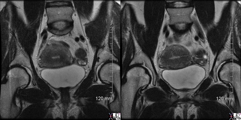
Thickened Junctional Zone |
| This T2 weighted MRI of a 41 year old female shows thickened junctional zone of the uterus measuring up to 12 mms characteristic of adenomyosis
83298c.81s uterus junctional zone thickened enlarged MRI T2 weighted Adenomyosis the uterus Courrtesy Ashey Davidoff MD copyright 2009 ectopic tissue |
|
Adenomyosis Thickened Junctional Zone Enlarged Uterus |
| This T2 weighted MRI of a 41 year old female shows thickened junctional zone (light maroon) of the uterus measuring up to 12 mms characteristic of adenomyosis
Courtesy Ashey Davidoff MD copyright 2010 |

|
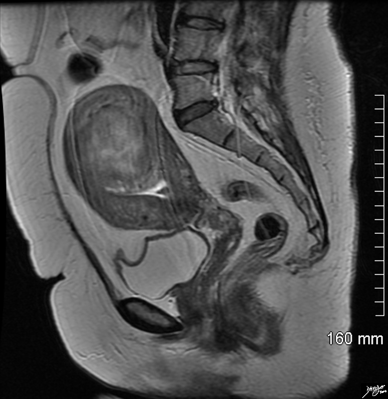
Deformity of the Endometrial CAvity Caused by an Intramural Fibroid |
|
In this sagittal view of the T1 weighted contrast enhanced study of the the uterus of a 46 year old female. The uterus roughly retains its pear shaped structure but the shape of the endometrial cavity is distinctly abnormal as if there is something pushing on it from above. This patient has a diffusely enhancing isointense leiomyoma that impinges on the endometrial cavity. Courtesy Ashley Davidoff MD Copyright 2010 All rights reserved 96577.8s |
|
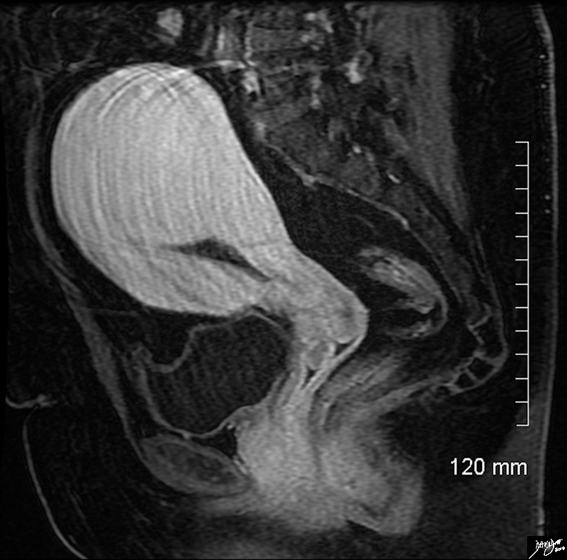
Large Intramural Fibroid Deforming the Stripe T2 Weighted Image |
|
In this sagittal view of the T2 weighted MRI of the uterus of a 46 year old female an almost 6cms slightly hyperintense fibroid is seen pushing on the endometrial cavity from above. This patient has a large fibroid aka leiomyoma that impinges on the endometrial cavity. Courtesy Ashley Davidoff MD Copyright 2010 All rights reserved 96570.8s |
|
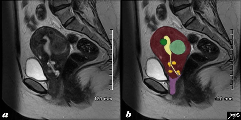
Fibroid Impingement on the Endometrium |
|
In this sagittal view of a T2 weighted MRI of the uterus of a 42 year old female with multiple fibroids two of which impinge on the endometrial cavity (the larger (light green) from posterior and the smaller (dark green) from anterior resulting in a sigmoid shaped cavity (yellow). In addition incidentally noted Nabothian cysts (orange) are seen within the cervix. Courtesy Ashley Davidoff MD Copyright 2010 All rights reserved 96531c02.8s |
Congenital Anomalies
|
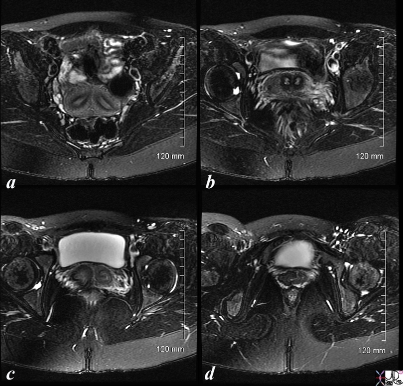
Two Hemiuteri, Two Cervices and One Vagina MRI T2 Weighted with FAt Saturation |
|
The MRI is from a 24F year old female with uterus didelphys The T2 weighted study in axial projection reveals 2 uterine corpuses (a) 2 cervices (b), vaginal septum in the upper 1/3 (c) but single distal vagina (d) Image Courtesy Ashley Davidoff MD Copyright 2010 83730c01L.8s |
|
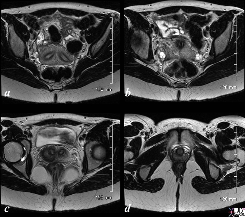
Two Hemiuteri, Two Cervices and One Vagina MRI T2 Weighted |
|
The MRI is from a 24F year old female with uterus didelphys The T2 weighted study in axial projection reveals 2 uterine corpuses (a) 2 cervices (b), vaginal septum in the upper 1/3 (c) but single distal vagina (d) Image Courtesy Ashley Davidoff MD Copyright 2010 83730c03L.8s |
References
Hamm, B Rosemarie Forstner, R, Beinder E MRI and CT of the female pelvis
Hricak, H, Carrington BM. MRI of the pelvis: a text atlas