Time and Aging
The Common Vein Copyright 2010
Introduction
In the fetus uterus is contained in abdominal cavity projecting beyond superior inlet of pelvis and cervix is considerably bigger that the body.
At puberty the uterus is pyriform in shape, and weighs from 14 to 17 gm. It has descended into the pelvis, the fundus being just below the level of the superior aperture of this cavity.
The position of the uterus in the adult is liable to considerable variation, depending chiefly on the condition of the bladder and rectum. When the bladder is empty the entire uterus is directed forward, and is at the same time bent on itself at the junction of the body and cervix, so that the body lies upon the bladder. As the latter fills, the uterus gradually becomes more and more erect, until with a fully distended bladder the fundus may be directed backward toward the sacrum.
During menstruation the organ is enlarged, more vascular, and its surfaces rounder; the external orifice is rounded, its labia swollen, and the lining membrane of the body thickened, softer, and of a darker color.
During pregnancy the uterus becomes enormously enlarged, and in the eighth month reaches the epigastric region. The increase in size is partly due to growth of preexisting muscle, and partly to development of new fibers.
After parturition the uterus nearly regains its usual size, weighing about 42 gm.; but its cavity is larger than in the virgin state, its vessels are tortuous, and its muscular layers are more defined; the external orifice which was round in nulliparous state may become slit like with one or more fissures.
In old age the uterus becomes atrophied, and paler and denser in texture; a more distinct constriction separates the body and cervix. The internal orifice is frequently, and the external orifice occasionally obliterated.
Season of the Uterus From Birth to Old Age |
|
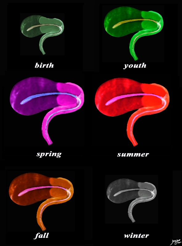
The sagittal diagram of the uterus and vagina through time shows an anteverted uterus at birth, through youth as it slowly matures and enlarges, entering spring as it reaches maturity, summer time when it bears fruit, and then through fall as it starts to involute in the postmenopausal period, and finally in its winter when it shrivels. In this view the uterus vagina, and the internal cavity starting in the endometrial cavity, coursing through the cervix and then into the collapsed vaginal cavity is exemplified. Courtesy Ashley Davidoff MD Copyright 2010 All rights reserved 96268b.51kc02Lh.8s |
The Adult Uterus
|
Non Gravid , 32 Week Pregnancy, and PostPartum Uterus |
|
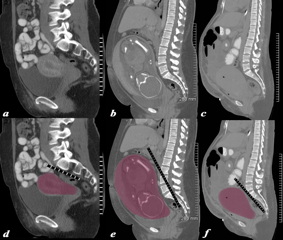
The series of CT scans from different patients are reconstructed in the sagittal plane to show the mature uterus in the non-gravid state (a,d) with a 32 week pregnancy (b,e), and in the postpartum, post cesarean section state (c,f). In the nongravid adult the uterus the craniocaudad span (c-c) measures about 9cms and the anteroposterior (A-P) dimension it measures 4.5cms The uterus containing the 32week pregnancy measures 24cms (c-c) by 16cms (A-P). In the post cesarean section patient the uterus measures 17cms (c-c) by 9cms (A-P). Courtesy Ashley Davidoff MD Copyright 2010 All rights reserved 78093c08.8s |
|
32 week Pregnancy |
|
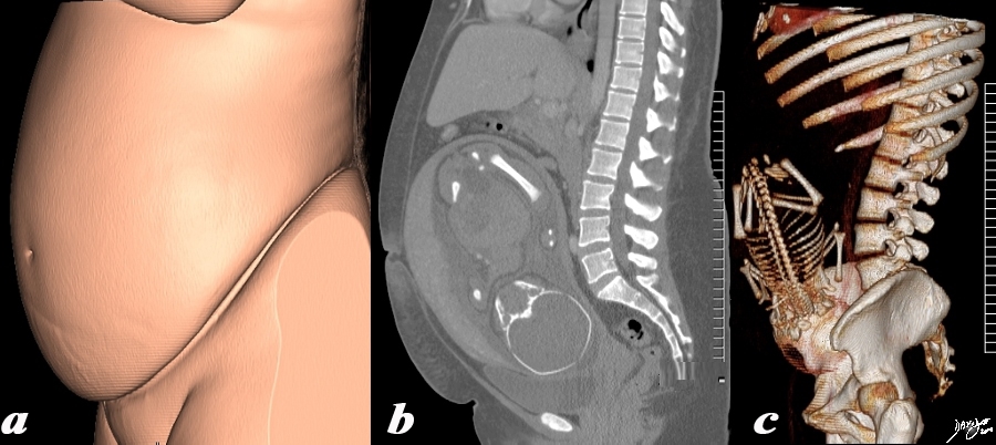
The 3 sagittally reconstructed images of the gravid uterus are from a normal 31 year woman carrying a 32 week gestation. They depict the outside appearance of the patient (a), 2 D sagittal view (b) and 3D sagittal view(c). Pregnancy is the main reason for living for the uterus and the opportunity to use it for this purpose is only a brief time in the long life of an adult female – but the species depends on this brief sojourn. Courtesy Ashley Davidoff MD Copyright 2010 All rights reserved 96332c.91s |
|
Twin Pregnancy – Pushing the Limits |
|
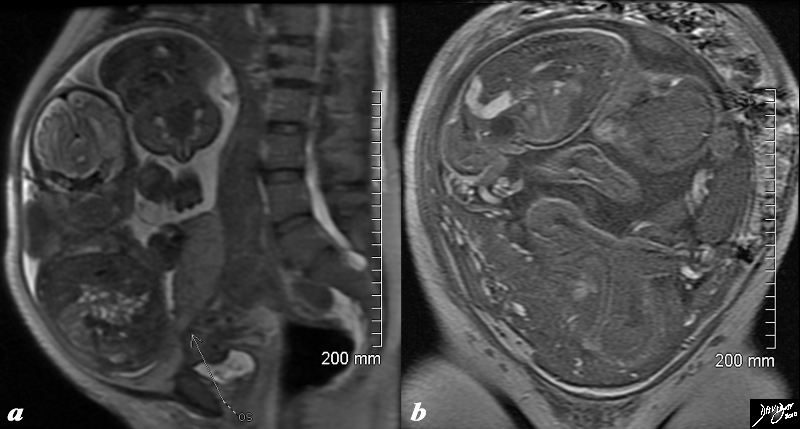
The T2 weighted MRI from a 44 year old patient with 32 week twin gestation in the sagittal plane (a) and in the coronal plane (b) revealing a uterus that measures 30cms in craniocaudad span, by 18cms A-P, by 26cms in the transverse plane. Courtesy Ashley Davidoff MD Copyright 2010 All rights reserved 88845c01.8s |
|
Post C Section and 18 Months Later |
|
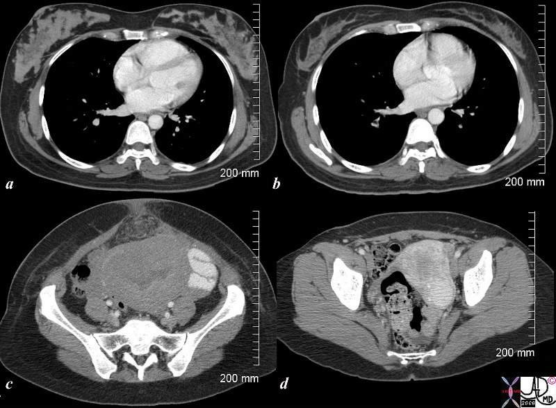
The CTscan is from a 26 year old female showing breasts and uterus in a post partum post cesarian section state (a,c) and then 18months later (c,d). The transverse dimension of the uterus in the post partum state is about 11cms, while 18months later is about 5cms. Her breasts in the post partum state are enlarged with prominent glandular tissue (a), and in the post partum state are reduced in size and glandular volume (b). Surgical footprints are noted in the subcutaneous tissue in c following her cesarian section. A cervical fibroid is suggested post partum image (d). Courtesy Ashley Davidoff MD Copyright 2010 All rights reserved 83354c.8s |

Post Partum Uterus
Then mean size of the uterus 24hours after delivery is 14X 7cms.(Garagiola)
The Uterus in Old Age
|
The Post menopausal Uterus 82 years old |
|
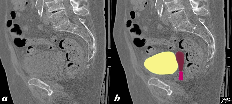
The sagittaly reconstructed CT is shown of an 82 year old female The craniocaudad span (c-c) is about 5cms and A-P dimension is 1.5cms. The endometrial stripe is is not visible. The uterus is buoyed and cushioned by partly filled bladder (yellow) Note the degenerative changes in the spine and the calcification of the distal abdominal aorta. Courtesy Ashley Davidoff MD Copyright 2010 All rights reserved 83564c04.8s |
Time Growth Cycles Aging
Menses |
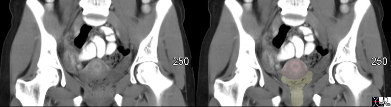
| 47670c01 uterus hyperemic endometrium soft tissue in vagina patient during menstruation menses normal physiology cycles time CTscan Davidoff MD |
|
Immediate Post Partum and 18 Months Later |
| 26 year old female presents 18 months after post c/section with gonadal vein thrombosis. Her breasts 18 months later show the glandular tissue is decreased compared to the immediate post partum period .
CTscan Courtesy Ashley DAvidoff copyright 2009 83354c.8s |