
The Common Vein Copyright 2010
Introduction
At Birth
The body of the uterus is similar in size to the cervix. Its overall size is 2.3-4.6 cms. (Nabaloff)
|
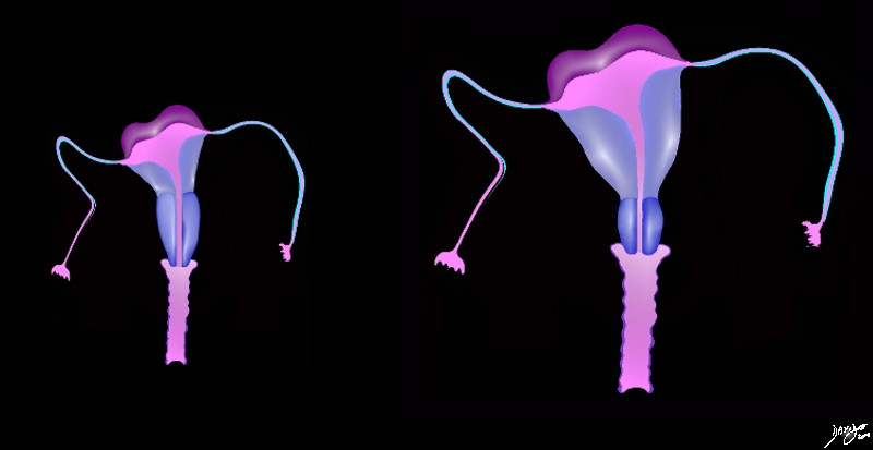
The Uterus at Birth and in the Adult |
|
This diagram in the coronal plane demonstrates the differences in size of the uterus at birth (left) and the mature, post menarche uterus (right). There is not only a difference in the absolute size but the ratio of the size body to the cervix changes as well. At birth the cervix is about the same size as the body of the uterus and post menarche the cervix is about 1/4 to 1/3 the size the body. Courtesy Ashley Davidoff MD Copyright 2010 All rights reserved 96266b15b01h02.83sc |
Prepubertal State
In virgin state the uterus is flattened antero-posteriorly and is pyriform in shape, with the apex directed downward and backward.
The prepubertal uterus should measure about 2-4.4cms in craniocaudad (c-c) span.
The Mature Uterus
The nulliparous uterus should measure about 6-8.5cms in c-c span by 2-4cms in A-P by 3-5cms in transverse.
The multiparous uterus should measure about 8-10.5cms in c-c span by 3-5cms in A-P by 4-6 cms in transverse.
|
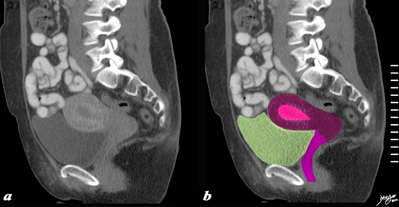
Normal Adult Uterus |
|
The sagittaly reconstructed CT shows an anteverted uterus buoyed and cushioned by partly filled bladder (yellow) In this sagittal view the adult uterus measures about 10cms in craniocaudad span and 5cms in A-P dimension and the endometrial stripe measures about 1cms. Courtesy Ashley Davidoff MD copyright 2010 all rights reserved 60385c03.81s |
|
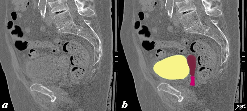
Normal Adult Uterus |
|
The normal sagittal view of the uterus is a reconstructed CT.. It demonstrates the normal size ratio of the body of the uterus to the cervix in the adult so that body(maroon) is about 2/3 the length and cervix about 1/3 (dark pink). The mature non gravid uterus measures about 8cm. in length, 4 cm. in A-P dimension, by nearly 6 cm. in transverse dimension; it weighs from about 35gms in the virgin state to up to 200 gms in the postpartum state, and has a correlative volume of between 75-200ccs.The myometrium measures between 1.5-2.5cms thick, and the endometrium measures from 1-10 mms and sometimes up to greater 10mms thick. Courtesy Ashley Davidoff MD Copyright 2010 All rights reserved 14707.2kb04i06.s.4ke06.8s |

The Gravid Uterus
|
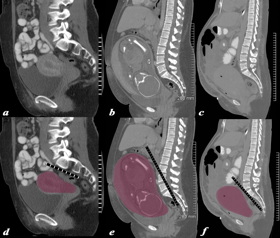
Non Gravid , 32 Week Pregnancy, and PostPartum Uterus |
|
The series of CT scans from different patients are reconstructed in the sagittal plane to show the mature uterus in the non-gravid state (a,d) with a 32 week pregnancy (b,e), and in the postpartum, post cesarean section state (c,f). In the nongravid adult the uterus the craniocaudad span (c-c) measures about 9cms and the anteroposterior (A-P) dimension it measures 4.5cms The uterus containing the 32week pregnancy measures 24cms (c-c) by 16cms (A-P). In the post cesarean section patient the uterus measures 17cms (c-c) by 9cms (A-P). Courtesy Ashley Davidoff MD Copyright 2010 All rights reserved 78093c08.8s |
|
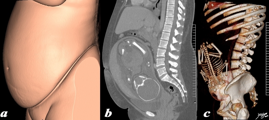
32 week Pregnancy |
|
The 3 sagittally reconstructed images of the gravid uterus are from a normal 31 year woman carrying a 32 week gestation. They depict the outside appearance of the patient (a), 2 D sagittal view (b) and 3D sagittal view(c). Pregnancy is the main reason for living for the uterus and the opportunity to use it for this purpose is only a brief time in the long life of an adult female – but the species depends on this brief sojourn. Courtesy Ashley Davidoff MD Copyright 2010 All rights reserved 96332c.91s |
|
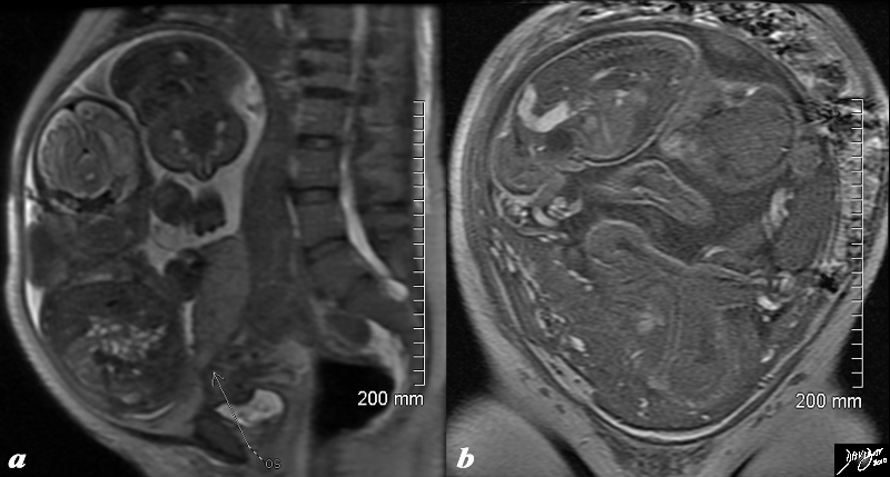
Twin Pregnancy – Pushing the Limits |
|
The T2 weighted MRI from a 44 year old patient with 32 week twin gestation in the sagittal plane (a) and in the coronal plane (b) revealing a uterus that measures 30cms in craniocaudad span, by 18cms A-P, by 26cms in the transverse plane. Courtesy Ashley Davidoff MD Copyright 2010 All rights reserved 88845c01.8s |
|
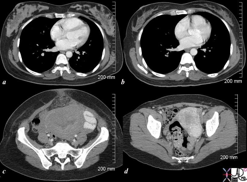
Post C Section and 18 Months Later |
|
The CTscan is from a 26 year old female showing breasts and uterus in a post partum post cesarian section state (a,c) and then 18months later (c,d). The transverse dimension of the uterus in the post partum state is about 11cms, while 18months later is about 5cms. Her breasts in the post partum state are enlarged with prominent glandular tissue (a), and in the post partum state are reduced in size and glandular volume (b). Surgical footprints are noted in the subcutaneous tissue in c following her cesarian section. A cervical fibroid is suggested post partum image (d). Courtesy Ashley Davidoff MD Copyright 2010 All rights reserved 83354c.8s |
Post Partum Uterus
Then mean size of the uterus 24hours after delivery is 14X 7cms.(Garagiola)
The Uterus in Old Age
|
The Post menopausal Uterus 82 years old |
|
The sagittaly reconstructed CT is shown of an 82 year old female The craniocaudad span (c-c) is about 5cms and A-P dimension is 1.5cms. The endometrial stripe is is not visible. The uterus is buoyed and cushioned by partly filled bladder (yellow) Note the degenerative changes in the spine and the calcification of the distal abdominal aorta. Courtesy Ashley Davidoff MD Copyright 2010 All rights reserved 83564c04.8s |

Adenomyosis |
| This T2 weighted MRI of a 41 year old female shows thickened junctional zone of the uterus measuring up to 12 mms characteristic of adenomyosis uterus junctional zone thickened enlarged MRI T2 weighted Adenomyosis the uterus
Courtesy Ashey Davidoff MD copyright 2009 14707c01.8s |