The Common Vein Copyright 2010
Introduction
|
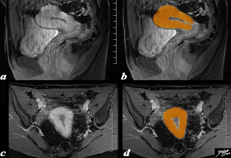
Pear Shape in both the Sagital and Coronal Planes |
|
The uterus is pear shaped as depicted in this overlay sagital (a,b) and coronal (c,d) T 1weighted enhanced MRI study Courtesy Ashley Davidoff MD Copyright 2010 96378c02.81s |
|
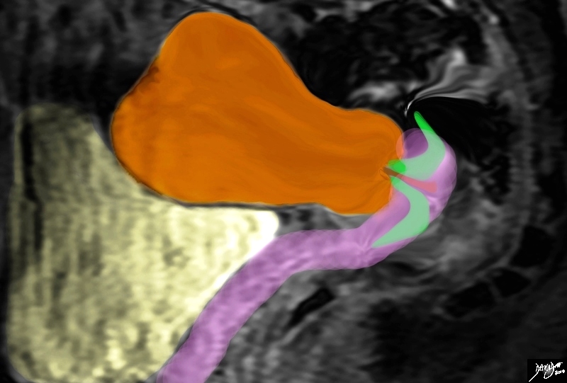
Sagital Plane |
|
The uterus is pear shaped as depicted in this overlay sagital MRI image Courtesy Ashley Davidoff MD Copyright 2010 6378c02kb05.8s |
|
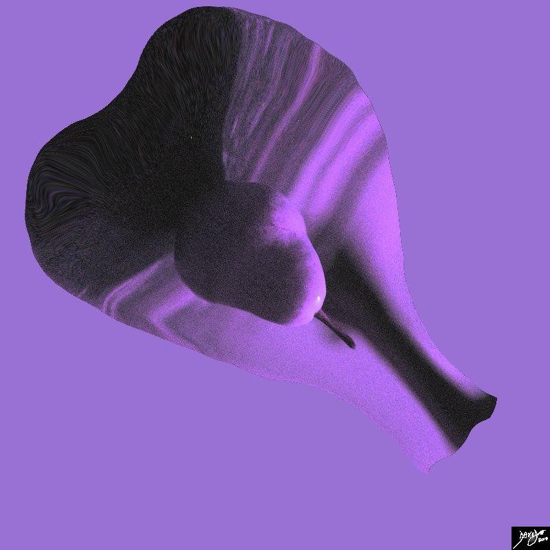
Shaped Like a Pear |
|
The uterus is shaped like a pear Courtesy Ashley Davidoff MD Copyright 2010 All rights reserved 85370pd07b.8s |
|
The Avocado – Similarities in Structure and Function |
|
The avocado pear has some interesting implications in the structure and function of the uterus. It is shaped like the eggplant and the pear. It takes between 6 and 12 months to grow an avocado from blossom to ripened fruit. The human gestational period is aboput 9 months uertine structure uterus food in the body Courtesy Ashley Davidoff MD Copyright 2010 All rights reserved 87836pb01d.8s |

|
The Uterus in the Sagittal Plane |
|
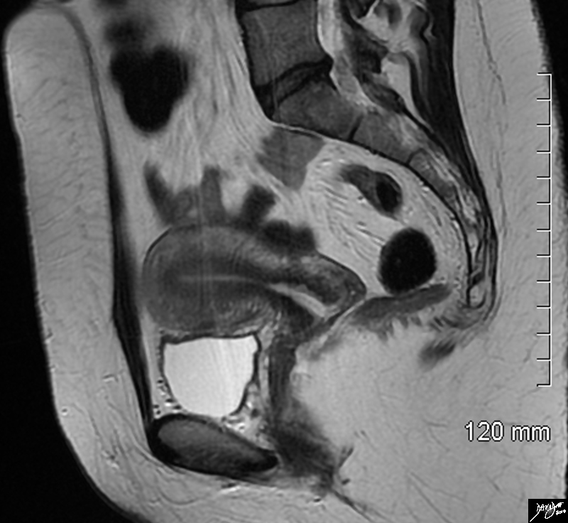
The sagittal STIR sequence from an MRI study shows the normal appearance of the uterus in a 34 year old female. The pear shaped form is exemplified together with the 3 parts of the uterus. The inner endometrium of intermediate signal, the junctional zone of low signal consisting of compacted smooth muscle with low water content, and the surrounding myometrium of smooth muscle with a higher water content. Courtesy Ashley Davidoff MD Copyright 2010 All rights reserved 96785.8s |
|
Axial Plane Coronal Projection |
|
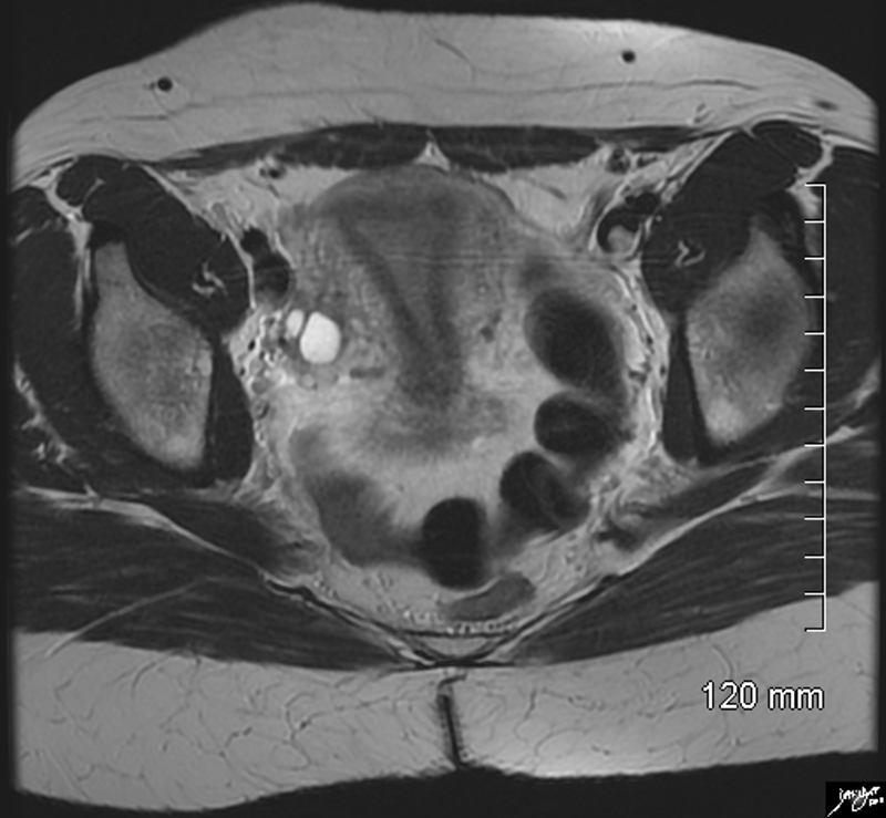
The axial STIR sequence from an MRI study shows the normal appearance of the uterus in a 34 year old female. Because the uterus is anteverted it lies flat so that in this instance the coronal plane of the uterus is demonstrated. The pear shaped form is exemplified together with the 3 parts of the uterus. The inner endometrium of intermediate signal, the junctional zone of low signal consisting of compacted smooth muscle with low water content, and the surrounding myometrium of smooth muscle with a higher water content. Two high signal cuysts are seen in the right ovary Courtesy Ashley Davidoff MD Copyright 2010 All rights reserved 96788.8s |
|
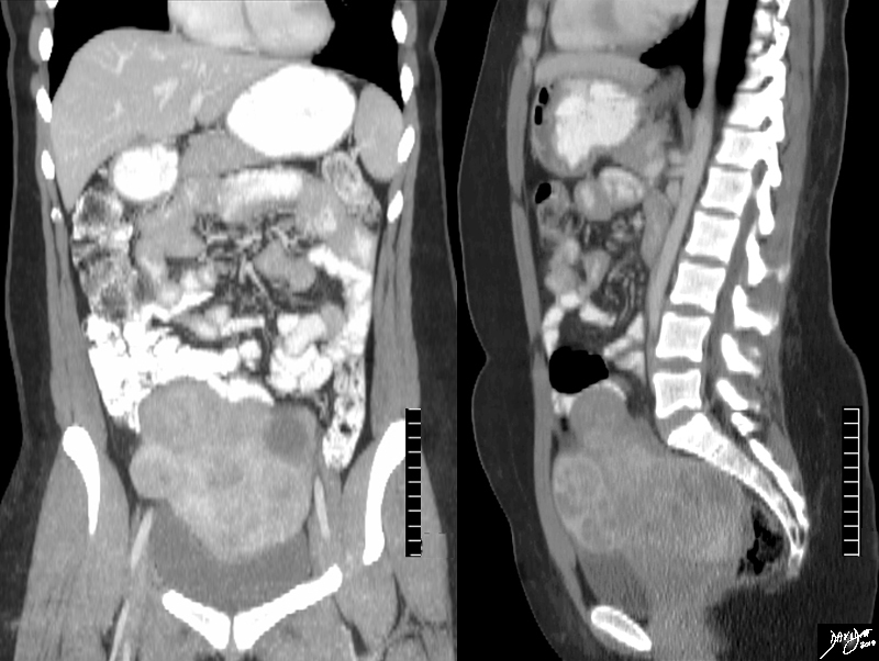
Large and Lobulated |
|
The CTscan is from a 31year old female, who presents with a pelvic mass.The study reveals a large uterus with multicentric fibroids and mild hydronephrosis The heterogeneous and lobular mass measures 17cms (c-c)by 10cms (A-P) by 12cms (transverse), and is consistent with a Courtesy Ashley Davidoff MD copyright 2009 all rights reserved 84362c.8s |