
The uterus, also known as the womb, is a hollow muscular organ that is part of the genitourinary system. It is characterized by its important role in the female reproductive system.
Structurally it is characterized by its pear shape and its unique muscular structure undergoes profound changes during different reproductive events. Though it is a pelvic structure it enlarges many times over to become an intrabdominal organ during pregnancy and returns to its prepregnancy state after child birth. Superiorly the gracile fallopian tubes open on each side into uterine cavity, and inferiorly it communicates with the vagina through the cervix. The urinary bladder lies anterior to it and rectum to posterior.
Functionally it provides a nutritional environment for a fertilized embryo to embed and accommodates a growing embryo until child birth.
Common diseases include alterations in the structure which can be congenital or acquired, benign or malignant tumors. Systemic disease especially infections can also affect uterus and uterine cavity.
Clinically disease usually manifests as infertility, recurrent abortions, menorrhagia, acute or chronic abdominal pain and urinary complaints.
Diagnosis is usually by detailed history and physical exams with imaging modalities like ultrasound,hysterosalpingography, hysteroscopy and diagnostic laparoscopy being most widely used.
Treatment options are guided by disease process and may include minimally invasive or open surgery.
|
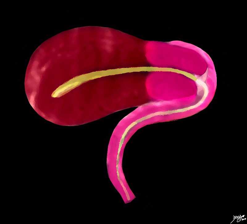
The Uterus and Vagina – Isolated The Pathway to Conception
|
|
The sagittal diagram of the uterus and vagina shows an anteverted uterus, which is the usual and normal position. In this view the uterus vagina, and the internal cavity starting in the endometrial cavity, coursing through the cervix and then into the collapsed vaginal cavity is exemplified. Courtesy Ashley Davidoff MD Copyright 2010 All rights reserved 96268b.51k.8s |
|
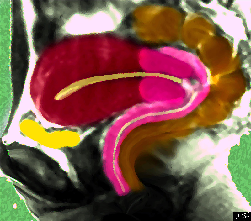
The Uterus Vagina and their Neighbors |
|
The sagittal diagram of the uterus shows the uterus surrounded by its neighbours, including the empty bladder (yellow) that lies anteriorly and inferiorly and the rectosigmoid colon that lies superiorly and posteriorly. Courtesy Ashley Davidoff MD copyright 2009 14707.2kb04i06.s.4k.8s |
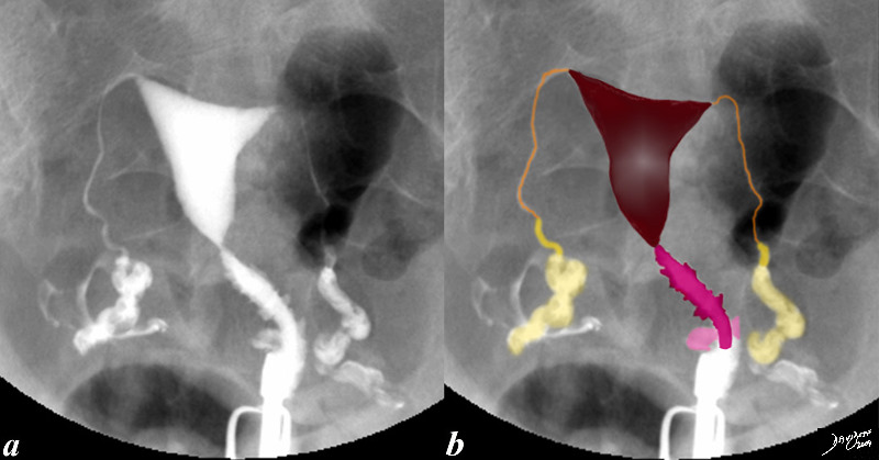
Normal Hysterosalpingogram |
|
This hysterosalpingogram is from a 26 year old female revealing a normal endometrial cavity and cervical canal. Note the irregular shape to the cervical canal while the endometrial lining is smooth. Note also the free spillage of contrast from the tubes into the peritoneal cavity (white contrast in b, indicating patent tubes. Courtesy Ashley Davidoff MD Copyright 2009 all rights reserved 83690c02.8s |