
The Common Vein Copyright 2010
Introduction
The uterus has three basic histological layers;
endometrium (aka mucosa) which is the inner layer abutting the lumen
myometrium which is the middle and thickest layer
serosa – the superficial layer

Basic Histological Layers – Body of the Uterus |
|
The wall of the uterus contains 3 basic layers; the mucosa, the muscularis and the serosa as demonstrated in this artistic rendition of the wall of the uterus in the premenstrual phase. Courtesy Ashley Davidoff MD Copyright 2010 All rights reserved 12047e04a03.8s |
The Endometrium (Mucosa)
The endometrium is a dynamic structure and in the reproductive female is turned over every thirty days.
The endometrium is subject to the influences of the hormonal changes of the menstrual cycle but only the endometrial component shows the changes.
It is divided into two zones; a thinner basal layer called the stratum basalis and a thick superficial functional layer called the stratum functionalis.
The stratum functionalis is considered a temporary tissue and since it comes and goes in 30 days it has a “unfinished” appearance. The stroma resembles embryonic tissue and does not have a characeristic appearance of a lamina propria.
The stratum basalis has a more permanent appearance characterized by mature appearing stromal tissue together with the deep tips of the glands
The epithelium is made of simple tubular glands that dip down like test tubes into the stroma. The epithelium contains ciliated columnar cells and secretory cells embedded in a highly cellular connective tissue with numerous lymphatics, vessels and uterine glands. The mucosa folds inwards to form simple tubular uterine glands that extend deep into the stroma.
The straoma consists of vessels lymphatics and connective tissue.
During the cycle both the epthelium and the stroma undergo significant change. At the time of menstruation the superficial functional layer which is sensitive to hormonal influences sloughs and the basal layer remains intact to enable regeneration during the next cycle.
The mucosa is continuous into the Fallopian tubes and opens into the to peritoneum. It also continues downstream into the vagina through external os.

Subparts of the Three Basic Layers |
|
The diagram typifies the histological appearance of the wall of the body of the uterus with other parts that are subdivisions of the 3 basic parts. The mucosa has two layers the superficial functional layer called the stratum functionalis and a deeper basal layer called the stratum basalis. The mucosa contains deep simple tubular glands embedded in a matrix (cream) The muscularis muscularis – (pink) consists of 3 layers; the subendometrial junctional zone organized in a circular fashion, and an outer myometrium which has two layers; The middle zone with criss cross pattern is the thickest and the thin circular outer zone. On the surface the thin serosa (mauve ) acts as a capsule for the uterus and a lining of the endometrial cavity. Courtesy Ashley Davidoff MD Copyright 2010 All rights reserved 12047e08c01.91s |
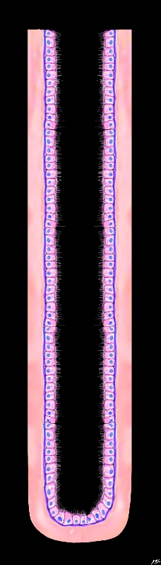
Simple Tubular Gland of the Endometrium |
|
The diagram reveals the histological appearance of the simple tubular gland of the endometrial lining of the uterus The ciliated columnar cells rest on a basement membrane (deep blue) which in turn are embedded in a stroma (pink) Courtesy Ashley Davidoff MD Copyright 2010 All rights reserved 32347f02.8s |
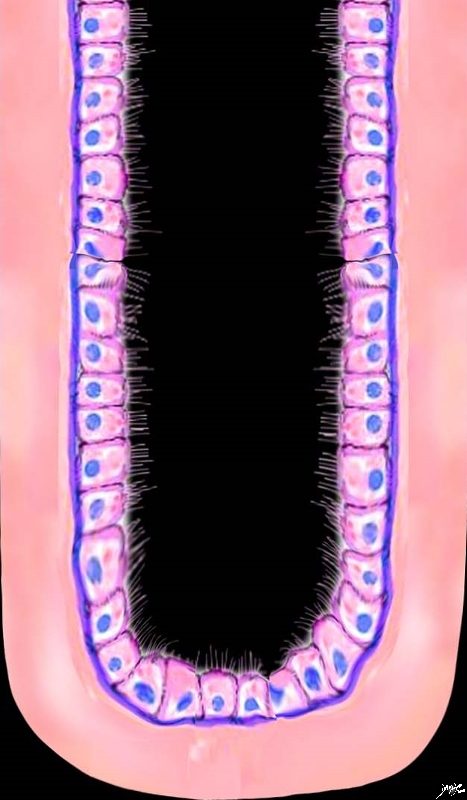
Appearance of the Cells – Magnified View |
|
The higher power diagram reveals the cytological appearance of the simple tubular gland of the endometrial lining of the uterus showing the ciliated columnar cells resting on the basement membrane (deep blue) which in turn are embedded in a stroma (pink) Courtesy Ashley Davidoff MD Copyright 2010 All rights reserved 32347f02.81s |
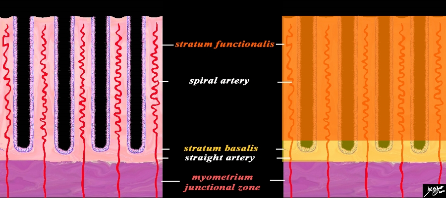
Functional Layer (Stratum Functionalis) Basal Layer (Stratum Basalis) and the Arteries Premenstrual Phase |
|
A closer view of the endometrium as seen in the premenstrual phase exemplified by the characteristic spiral (helical) arteries running in the stroma (pink) between the simple tubular test tube shaped glands (purple) The spiral arteries supply the functional layer (stratum functionalis deep orange) and the straight arteries supply the basal layer (stratum basalis- light orange). Beneath the basal layer is the myometrium- Subendometrial smooth muscle or junctional zone (dark pink). Courtesy Ashley Davidoff MD Copyright 2010 All rights reserved 32347f08cL.9s |

Distribution of the Arterial Supply Histologic Level |
|
This diagram exemplifies the position the arteries in the epithelial and muscular layers. The spiral arteries run in the stratum functionalis, the straight arteries run in the stratum basalis, while the radial arteries and the arcuate arteries run in the muscularis. Courtesy Ashley Davidoff MD Copyright 2010 All rights reserved 12047e04b04L02.8s |
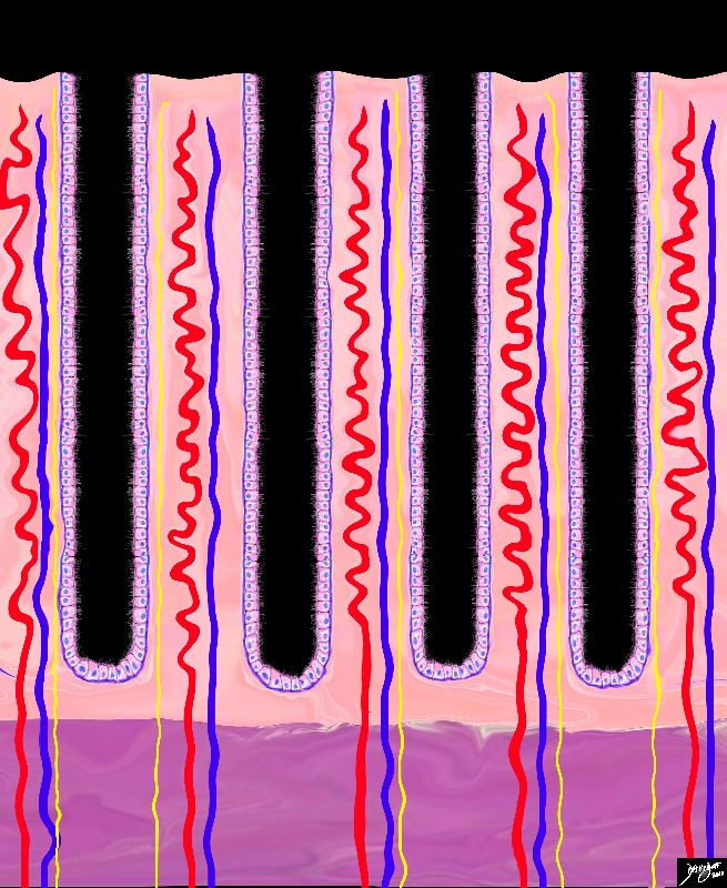
Arteries Veins and Lymphatics in the Stroma |
|
This diagram of the mucosa shows some of the other vessels in the stroma including the veins (blue), the lymphatics (yellow) andthe already described spiral and straight arteries (red) Courtesy Ashley Davidoff MD Copyright 2010 All rights reserved 32347f11.8s |
Cyclical Changes of the Endometrium
The three major phases of the cycle that manifest in the endometrium are the proliferative (follicular), secretory (luteal), and menstrual phases.
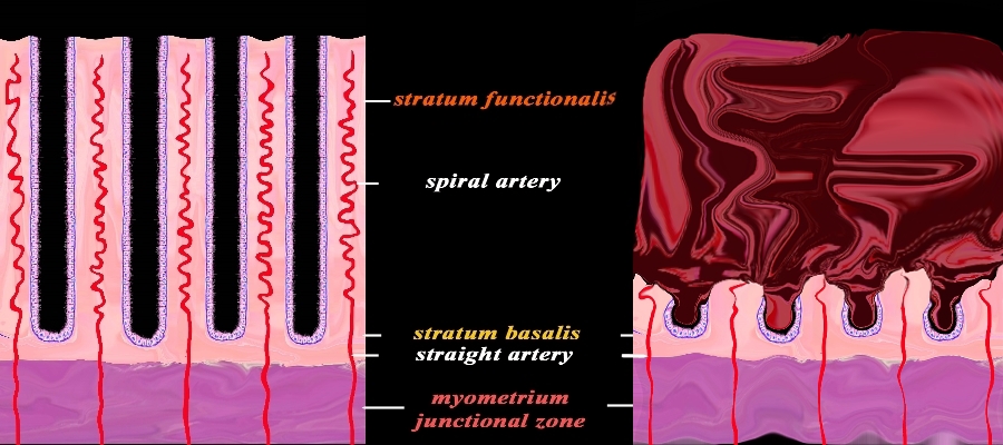
Premenstrual (left) and Active Menstruation (right) |
|
The diagram reflects the premenstrual endometrium (left) and the post menstrual endometrium right, revealing the necrosis of the functional layer with sloughing and hemorrhage. The basal layer with the staright arteries and a small portion of the spiral artery remains intact. The hemorrhage is controlled by spasm of the arteries and contraction of the myometrium. Courtesy Ashley Davidoff MD Copyright 2010 All rights reserved 32347f08cL.92s |
In the cervix the mucous membrane is sharply differentiated from that of the uterine cavity. The cervical mucosa is thrown into numerous oblique ridges, which diverge from an anterior and posterior longitudinal raphé. In the upper two-thirds of the canal, the mucous membrane is provided with numerous deep glandular follicles, which secrete a clear viscid alkaline mucus; and, in addition, extending through the whole length of the canal is a variable number of little cysts, presumably follicles which have become occluded and distended with retained secretion. These are called as ovula nabothi. The upper two thirds of cervix is ciliated cylindrical epithelium below which it gradually transforms into stratified squamous epithelium. On the vaginal surface of cervix the epithelium is similar to the vaginal epithelium.
The Muscular Layer
The muscular layer forms the bulk of uterus. It is thick in fundus and opposite the middle part of the body and thin at entry point of Fallopian tubes. There are three component muscular layers; external, middle, and internal.
The external layer consists of fibers which pass transversely across the fundus, and, converging at each lateral angle of the uterus. They continue on to the Fallopian tube, the round ligament, and the ligament of the ovary. Some of the fibers pass on each side into the broad ligament, and some continuebackward from the cervix into the sacrouterine ligaments.
The middle layer has no regular arrangement of the fibres they are disposed longitudinally, obliquely and transversely. This layer has largest number of blood vessels. After parturition these layers act as living ligatures and limit blood loss .
The internal or deep layer is like muscular mucosa ; the fibres are arranged in circular fashion like two hollow cones with apices at uterine tube orifices and bases intermingling in the middle of the body. The fibres at the internal os are arranged in circular fashion like a sphincter.
During pregnancy the muscles undergo both hyperplasia (more snmooth muscle cells) and hypertrophy (larger muscle cells).
The Serosa
The serosa does not cover the uterus completely. It is derived from the peritoneum; it invests the fundus and the whole of the posterior intestinal surface of the uterus; but covers the vesical surface only as far as the junction of the body and cervix. In the lower fourth of the posterior surface, the serosa is loosely attached to the uterus separated by connective tissus. the reflection of intestinal peritoneum from uterus to intestine forms pouch of Douglas. This is the area where pelvic collection such as abscess can accumulate and can be drained vaginally.
References
Histology at Southern Illinois University School of Medicine
Noe, M, Kunz, G, Herbertz, M, Mall, G, Leyendecker, G. The cyclic pattern of the immunocytochemical expression of oestrogen and progesterone receptors in human myometrial and endometrial layers: characterization of the endometrial–subendometrial unit Oxford Journals Human Reproduction Volume 4 Issue pp 190-197