The Common Vein
Copyright 2010
Definition
Uterine obstruction is an anatomic abnormality of the uterus.
Causes of uterine obstruction include congenital abnormalities of the uterus, scarring of the cervix from surgery or radiation and tumor obstructing the endocervical canal.
The result is a uterine cavity which does not connect to the lower reproductive tract.
The structural changes depend on the cause of obstruction. In patients with congenital uterine anomalies, an abnormality in fusion or development of the mullerian ducts results in a uterine body with no outlet to the vagina at birth. After surgery or radiation involving the cervix, inflammation and scarring can occur causing cervical stenosis and eventually complete obstruction. Uterine, cervical or vaginal tumors may grow to obstruct the endocervical canal.
The functional changes of uterine obstruction are characterized by blood collecting in the uterine cavity (hematometria) without an outlet at menses.
As a result, women with uterine obstruction present with amenorrhea and cyclic abdominal pain. Infection can occur in the blood filled uterine cavity (pyometria) leading to fever and bacteremia.
Diagnosis is based on physical exam and imaging with ultrasound.
Treatment depends on the cause, but always includes relief of the obstruction. In pyometra, drainage of the infected blood must be accompanied by treatment with antibiotics.
Cervical Stenosis
|
Cervical Stenosis and Metrorrhagia |
|
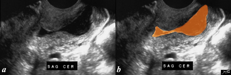
The transvaginal ultrasound is from a 50 year old perimenopausal female with metrorhagia. The uterine cavity and cervical cavity are filled with fluid, and soft tissue elements are identified in the expanded cervical canal. The findings are consistent with cervical stenosis, burt the cause of the metrirhagia is not obvious. The stenosis was relieved and follow up ultrasound showed resolution. No cervical mass was identified. Courtesy Ashley Davidoff MD copyright 2010 all rights reserved 85921c04.8s |
Polyp as a Cause of Obstruction Presumed Benign Disease |
|
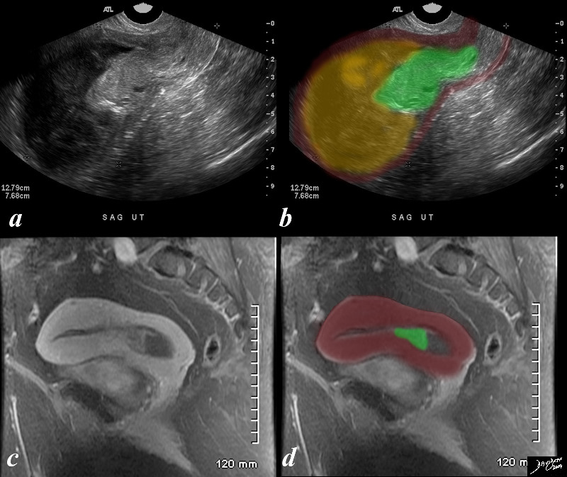
The ultrasound and MRIscan are from a 67 year old female, with an LMP of 10 years ago and with gynecologic history G9 and current history of bleeding for one month, a fever and low hematocrit. She has not had follow up gynecological examination for many years. The studies reveal a mass in the lower uterine segment and an obstructed endometrial cavity tha contains complex material which is presumed combination of blood and pus. ie hematometria and pyometria. The ultrasound is far more impressive than the MRI in that the mass (green) looks more impressive in size and there is prominent upstream accumulation of of complex material and fluids. The MRI shows a enhancing lesion arising from the endometrial mucosa, and at this time the fundal portion is relatively decompressed. The mass is presumably a benign polyp since her subsequent history to the hospital revealed only follow up for chronic renal failure and no pathological report of a malignancy has been n noted. Courtesy Ashley Davidoff MD copyright 2009 all rights reserved 84127c01.8s |
Cervical Cancer
|
Enlarged and Obstructed Uterus – Cancer of the Cervix |
| 46 year old female with obstructed uterus and fluid filled endometrial cavity with pockets of air caused by cervical carcinoma complicated by pyometria
Courtesy Ashley Davidoff MD Copyright 2009 all rights reserved 83648c02.8s |
|
Carcinoma of the Cervix in a Bicornuate Uterus Complicated by Obstruction |
| 74 year old female with bicornuate uterus and dilated endometrial cavities Diagnosis is carcinoma of the cervix with obstruction . The myometrium is overlaid in dark pink, and the endometrial cavity is a heterogeneous orange consisting of both fluid and soft tissue elements.
Incidental note is made of gastroesophageal reflux Courtesy Ashley Davidoff MD copyright 2009 all rights reserved 83438c01.8s |
|
Cervical Carcinoma Pre-obstructive |
|
31 year female with a mass in the lower uterine segment of the uterus (green overlay) There is almost total occlusion of the canal (yellow) There are small vessels feeding the tumor (red). The patient subsequently obstructed the endometrial cavity and became secondarily infected. Courtesy Ashley Davidoff Copyright 2009 all rights reserved 83474c01.8s |
|
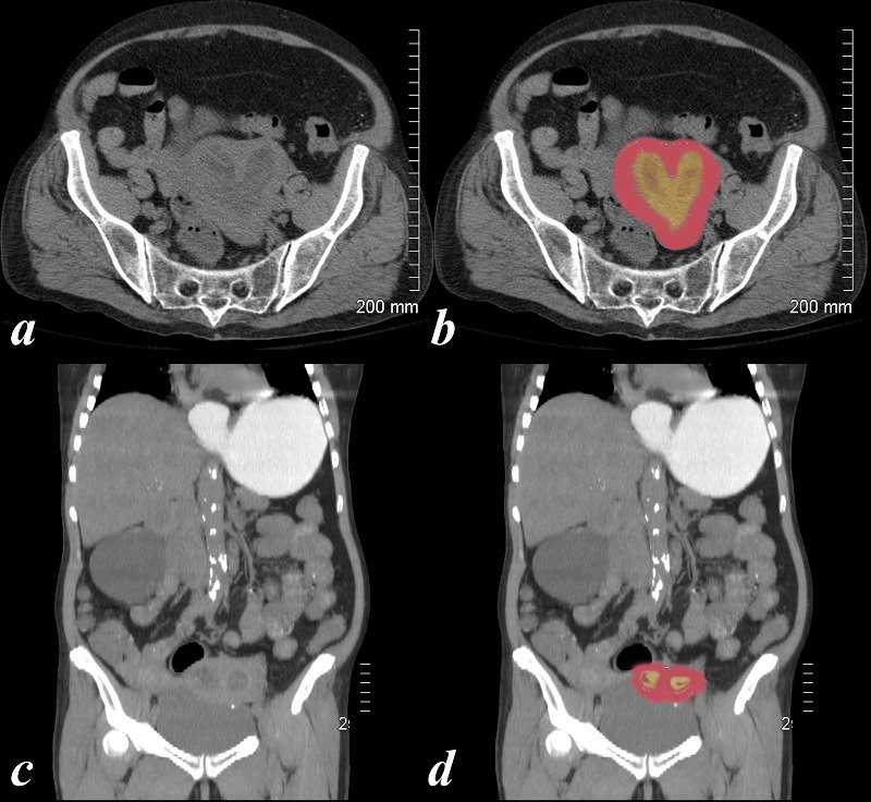
Endometrial Carcinoma |
|
The axial CTscan is from a 31 year old female with carcinoma. The images show an expanded irregular endometrial cavity (yellow) that contains air (black), soft tissue (green), and fluid (yellow), uterus with an enhancing filling defect (green overlay in b). The mass could arise from the cervix based on its position or from the uterus. The former is more likely based on the patients age.. A solid appearing mass is seen in the right adnexa (dark green) representing either a solid mass in the ovary or a enlarged iliac node. These findings are consistent cervical carcinoma or less likely endometrial with secondary obstruction and pyometria. 83484c02.8s Courtesy Ashley Davidoff Copyright 2010 all rights reserved air in endometrial cavity infection |
|
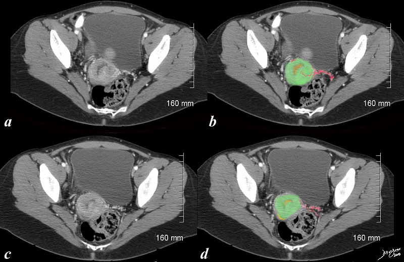
Obstruction and Infection |
|
The axial CTscan is from a 46 year old female with cervical carcinoma. The images show an expanded irregular endometrial cavity (yellow) that contains air (black), thickened irregular cervix (soft tissue green), and fluid (yellow). These findings are consistent cervical carcinoma or less likely endometrial with secondary obstruction and pyometria. 83682c01.8s Ashley Davidoff MD Copyright 2009 all rights reserved |
|
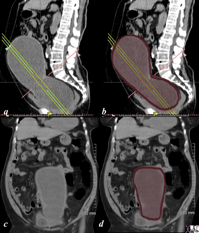
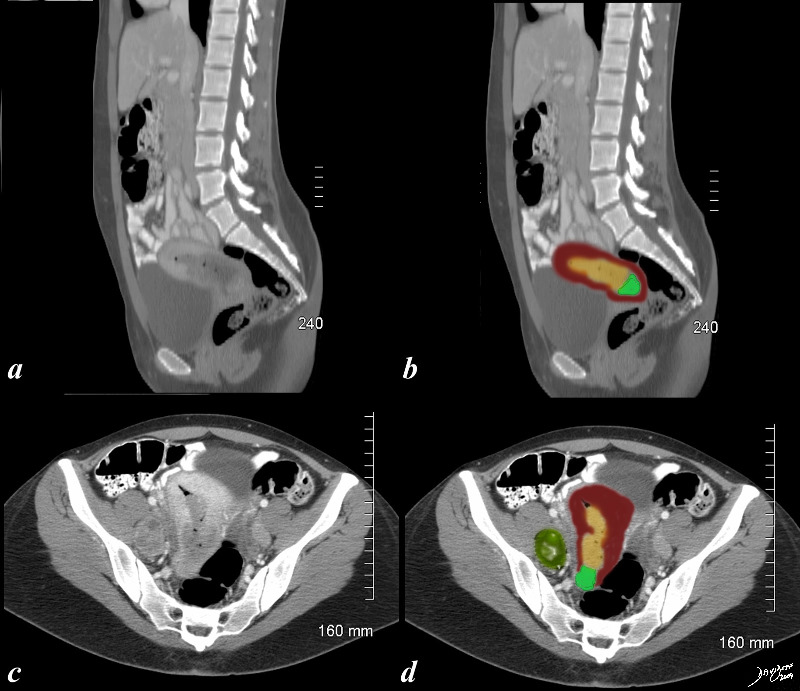
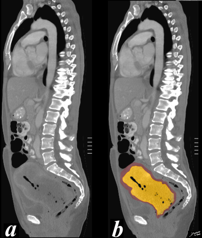
Obstructed Uterus with Pyometria |
| 46 year old female with obstructed uterus and fluid filled endometrial cavity with pockets of air caused by cervical carcinoma complicated by pyometria.
Courtesy Ashley Davidoff MD Copyright 2009 all rights reserved 83682c01b.8s |
|
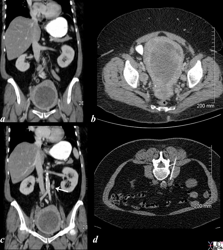
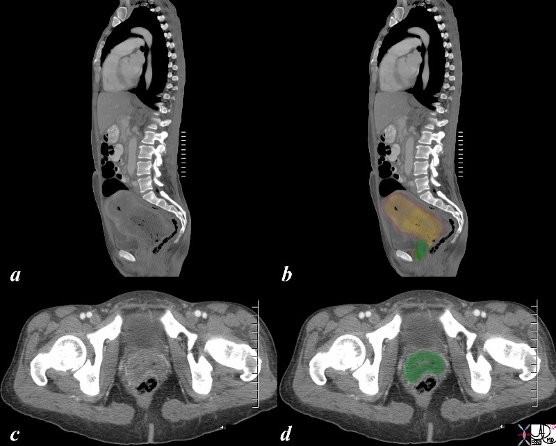
Metastatic Disease to the Para-Aortic Nodes |
| 61 year old female with enlarged endometrial cavity and cervical mass consistent with cervical carcinoma complicared by obstruction of the endometrial cavity. The cavity is filled with complex material probably from a combination of secretions, fluid, blood and tumor. Lymph nodes in the left paraaortic area (c, white arrow) were biopsied under CT guidance (d) and were positive for cervical cancer.
Courtesy Ashley DAvidoff copyright 2009 all rights reserved 85306c01.8s |