Copyright 2010
Introduction
Hyperplasia
|
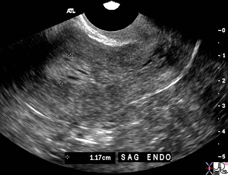
Thick Heterogeneous Endometrium |
|
The transvaginal ultrasound is from a 60 year old female who presents with spotting Ultrasound reveals a heterogeneous endometrial stripe consistent with endometrial hyperplasia though endometrial carcinoma is a possibility. Malignant neoplasia is a a les likely possibility Courtesy Ashley DAvidoff MD copyright 2010 83301.81s |
Carcinoma
|
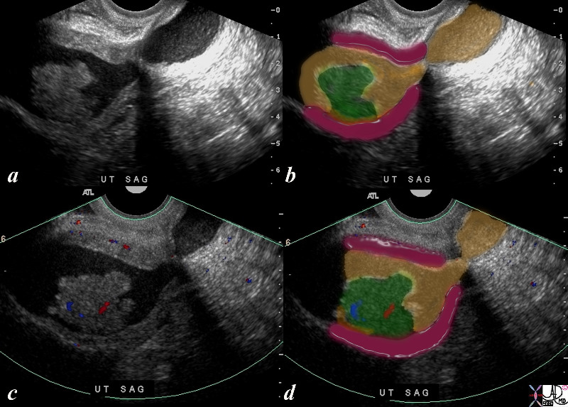
Endometrial Carcinoma and Uterine Obstruction |
|
The ultrasound is from a 70 year old post menopausal female who presents with an enlarged uterus. The endometrial stripe is enlarged and is filled with fluid and an enhancing soft tissue mass consistent with an endometrial carcinoma. Note blood flow as depicted by Doppler exam (c) characterizing the soft tissue as tumor rather than a clot. Courtesy Ashley Davidoff MD copyright 2009 all rights reserved 86206c.8s |
|
Focal Endometrial Thickening |
|
The coroanal CTreformatted CTscan images are from a 46 year old female with endometrial carcinoma. The images show an focal area of soft tissue thickening (green), on the superior surface (b) and rightward (green d) of the endometrial cavity (orange) . 83494c01.8s Courtesy Ashley Davidoff Copyright 2010 all rights reserved |

|
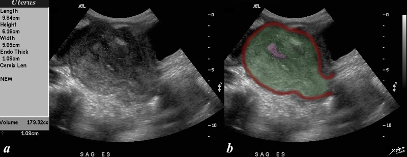
Endometrial Cavity Expanded with Tumor |
|
The ultrasound of a 58 year female with expanded endometrial cavity is shown. The cavity is filled with heterogenous material (green) representing a combination of tumor, fluid necrotic material and blood. Pathology showed adenocarcinoma, endometriod type, grade I/III with tumor necrosis. In this iamge a small portion of normal appearing endometrium is shown in pink. Tumor involvement with the right ureter and secondary hydronephrosis of the right kidney is present. Courtesy Ashley Davidoff Copyright 2009 all rights reserved 83510c03.8s |
|
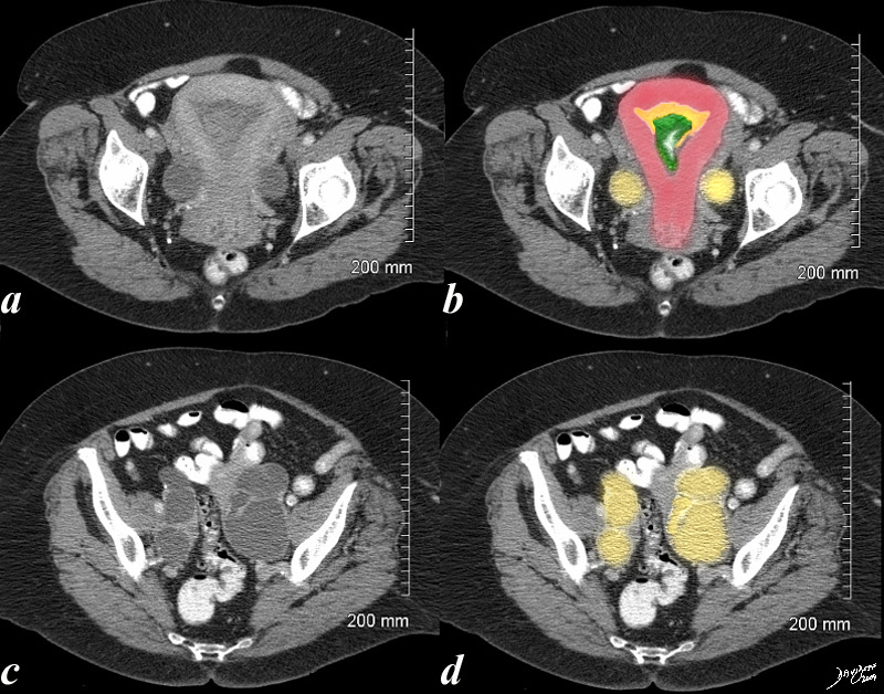
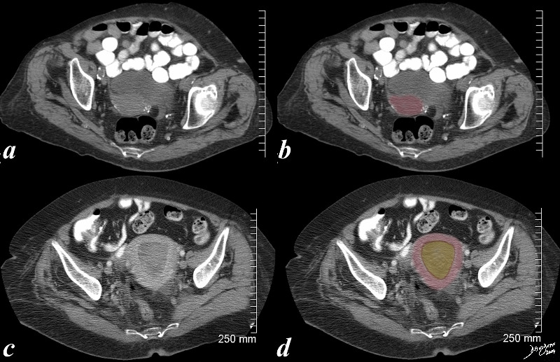
Assessing the Large Uterus in the Elderly |
|
The first CTscan (a,b) is from an 86 year old patient with a small but normal post menopausal uterus showing characteristic vascular calcification. The second CT (c,d) is from an 83F year old patient with distended endometrial cavity (orange in d) with known uterine carcinoma Courtesy Ashley Davidoff MD copyright 2009 all rights reserved 83731c01.8S |
|
Endometrial Carcinoma |
|
The axial CTscan is from a 31 year old female with carcinoma. The images show an expanded irregular endometrial cavity (yellow) that contains air (black), soft tissue (green), and fluid (yellow), uterus with an enhancing filling defect (green overlay in b). The mass could arise from the cervix based on its position or from the uterus. The former is more likely based on the patients age.. A solid appearing mass is seen in the right adnexa (dark green) representing either a solid mass in the ovary or a enlarged iliac node. These findings are consistent cervical carcinoma or less likely endometrial with secondary obstruction and pyometria. 83484c02.8s Courtesy Ashley Davidoff Copyright 2010 all rights reserved air in endometrial cavity infection |
|
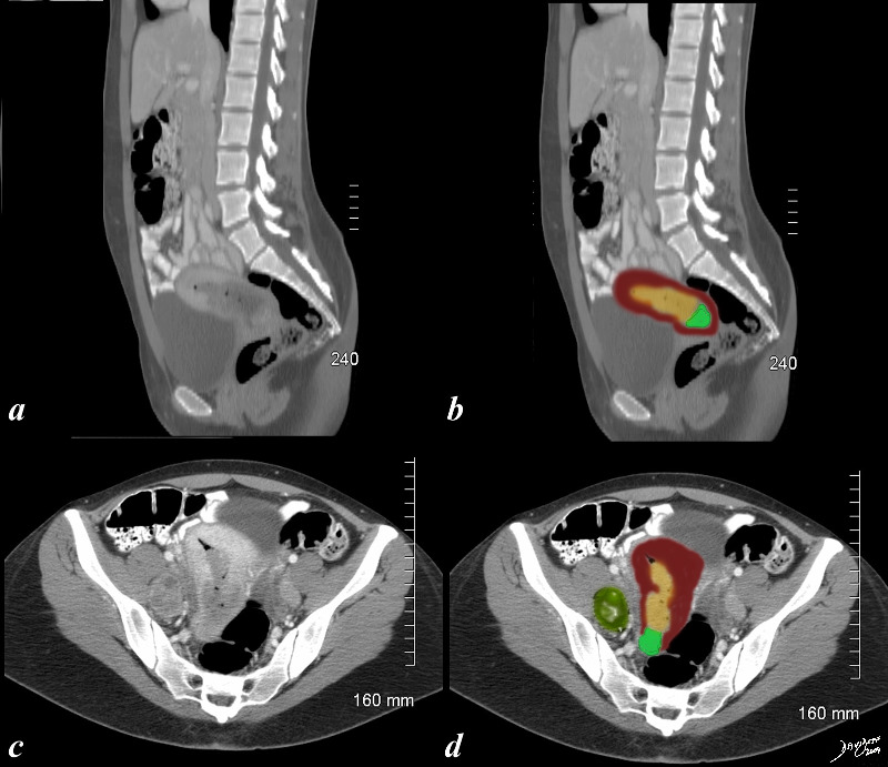
Endometrial Carcinoma with Hydrosalpinx |
|
This 70 year old female presents with pelvic discomfort. The CT shows an endometrium (orange in b) filled with complex soft tissue in the endometrial cavity (green in b) and the Fallopian tubes are distended with fluid (yellow in b and d) caused by the obstructing carcinoma. The uterus is enlarged Courtesy Ashley Davidoff MD copyright 2009 all rights reserved 48414c02.8s |