
AssThe Common Vein Copyright 2010
Identifying Uterine Disease and and Local Complications
|
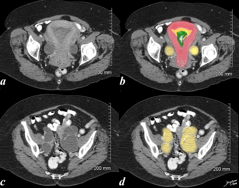
Enlarged Uterus Endometrial Carcinoma with Hydrosalpinx |
|
This 70 year old female presents with pelvic discomfort. The CT shows an endometrium (orange in b) filled with complex soft tissue in the endometrial cavity (green in b) and the Fallopian tubes are distended with fluid (yellow in b and d) caused by the obstructing carcinoma. The uterus is enlarged Courtesy Ashley Davidoff MD copyright 2009 all rights reserved 48414c02.8s |
Ability to Characterize Small Bubbles of Air and Incriminate Anaerobic Infection
|
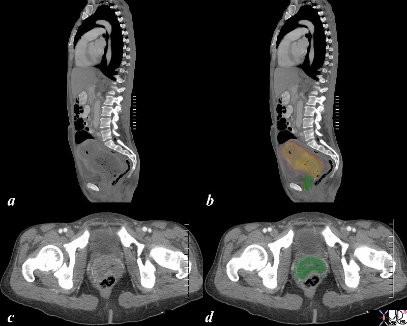
Obstruction and Infection |
|
The axial CTscan is from a 46 year old female with cervical carcinoma. The images show an expanded irregular endometrial cavity (yellow) that contains air (black), thickened irregular cervix (soft tissue green), and fluid (yellow). These findings are consistent cervical carcinoma or less likely endometrial with secondary obstruction and pyometria. 83682c01.8s Ashley Davidoff MD Copyright 2009 all rights reserved |
|
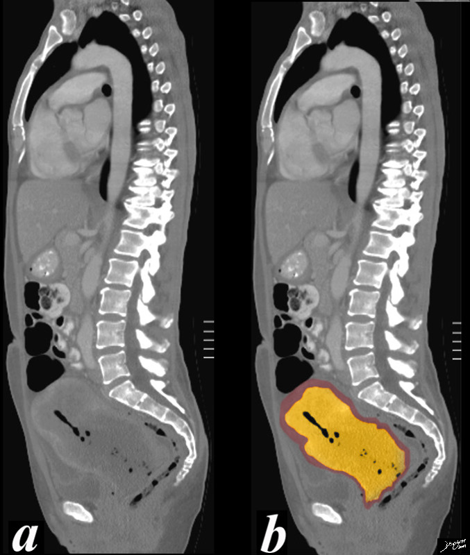
Obstructed Uterus with Pyometria |
| 46 year old female with obstructed uterus and fluid filled endometrial cavity with pockets of air caused by cervical carcinoma complicated by pyometria.
Courtesy Ashley Davidoff MD Copyright 2009 all rights reserved 83682c01b.8s |
Metastases
|
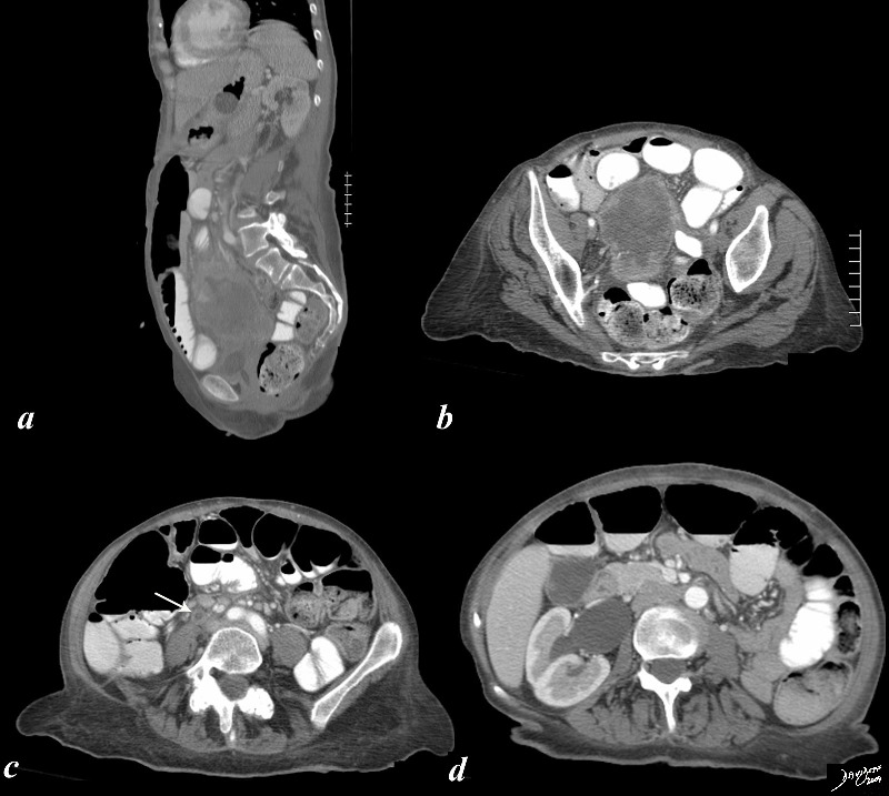
Metastasis to the Right Ureter Causing Hydronephrosis |
|
The CTscan of a 58 year female with expanded endometrial cavity is shown. (a,b) The cavity is filled with heterogenous material (green) representing a combination of tumor, fluid necrotic material and blood. Pathology showed adenocarcinoma, endometriod type, grade I/III with tumor necrosis. Tumor involvement of the right ureter (arrow in c) and secondary hydronephrosis of the right kidney is present. (d) Courtesy Ashley Davidoff Copyright 2009 all rights reserved 83510c07.8s |
Beyond the Pelvis
|
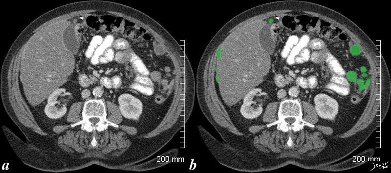
Peritoneal Metastases |
|
79 year female with transperitoneal metastasis from endometrial carcinoma overlaid in green. Fatty liver is present. Courtesy Ashley Davidoff Copyright 2009 all rights reserved 83526c.8s |
CT Guided Biopsy
|
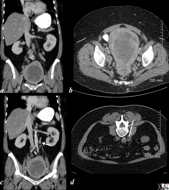
Regional Nodes |
|
61 year old female with enlarged endometrial cavity and cervical mass consistent with cervical carcinoma complicared by obstruction of the endometrial cavity. The cavity is filled with complex material probably from a combination of secretions, fluid, blood and tumor. Lymph nodes in the left paraaortic area (c, white arrow) were biopsied under CT guidance (d) and were positive for cervical cancer. Courtesy Ashley DAvidoff copyright 2009 all rights reserved 85306c01.8s |
—
References
Kaura, H, Loyera E M, Minamib, M Charnsangaveja C Patterns of uterine enhancement with helical CT European Journal of Radiology Volume 28, Issue 3, Pages 250-255 (October 1998)