
Copyright 2008
Definition
Uterine didelphys is a congenital anomaly of the uterus caused by failure of fusion of the mullerian ducts during embryonic development.
The result is a woman born with two separate uterine bodies.
The structural changes are characterized by two uterine horns, each connecting a separate cervix to a single fallopian tube. The vaginal cavity leading to the cervices is sometimes divided by a septum.
The functional change is variable, but can be characterized by limitation of fetal growth during pregnancy. The vaginal septum may also interfere with sexual intercourse.
Women typically present clinically with pain during intercourse (dyspareunia). However, uterine didelphys may also manifest with complications during pregnancy including infertility, pregnancy loss or preterm labor. Women with uterine didelphys are more likely to require cesarean section for delivery.
Imaging modalities used to evaluate uterine abnormalities include sonohysterography, hysterosalpingography, and MRI.
Diagnosis is made with physical exam combined with findings on imaging.
Treatment is limited to resection of the vaginal septum to decrease dyspareunia. In some studies, resection of the vaginal septum also improved fertility. Procedures to fuse the uterine bodies or remove a uterine body have not proven beneficial and are not typically performed for women with uterine didelphys.
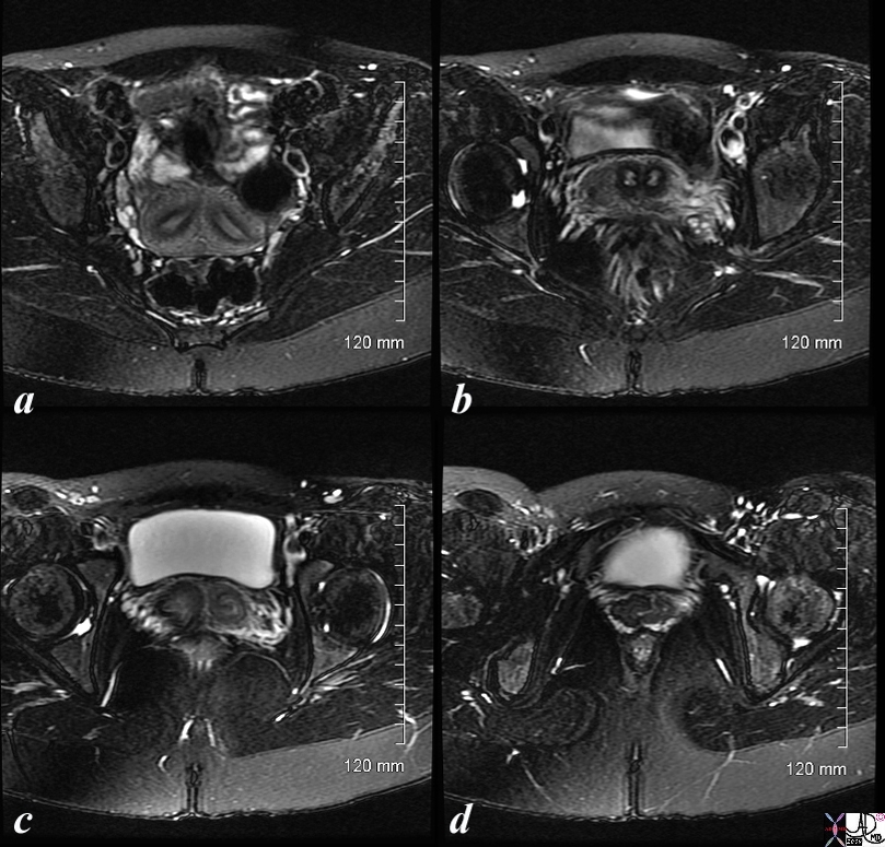
Two Hemiuteri, Two Cervices and One Vagina MRI T2 Weighted with FAt Saturation |
|
The MRI is from a 24F year old female with uterus didelphys The T2 weighted study in axial projection reveals 2 uterine corpuses (a) 2 cervices (b), vaginal septum in the upper 1/3 (c) but single distal vagina (d) Image Courtesy Ashley Davidoff MD Copyright 2010 83730c01L.8s |
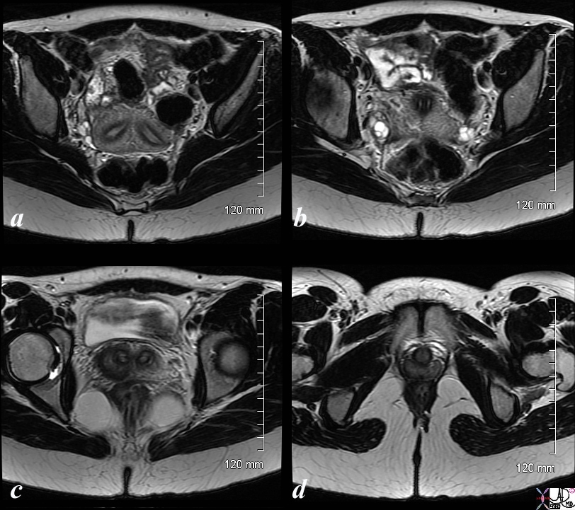
Two Hemiuteri, Two Cervices and One Vagina MRI T2 Weighted |
|
The MRI is from a 24F year old female with uterus didelphys The T2 weighted study in axial projection reveals 2 uterine corpuses (a) 2 cervices (b), vaginal septum in the upper 1/3 (c) but single distal vagina (d) Image Courtesy Ashley Davidoff MD Copyright 2010 83730c03L.8s |
Accounts for 5% of uterine anomaliescaused by nearly complete failure of fusion of te mullerian ducts
Thre are two hemiuteri and two cervices
Longitudinal septum is seen in 75%
There is no communication between two endometrial cavities unlike bicornuate where there are
Clinically infertitlity/hematrometrocolpos/dysmennorhea/endometriosis/pelvic adhesions
Rx surgery is controversial