
Copyright 2008
Definition
The Fallopian tubes (aka salpinges, oviducts) are a pair of tubular structures that are part of the female reproductive system and also part of the genitourinary system. They are characterized by their delicate and gracile nature and functional importance in fertilization.
Structurally they are usually about 10cms long (range 7-14cms), and are funnel shaped. The widest portion of the funnel is situated laterally, is called the infundibulum, and is open to the peritoneal cavity. The fimbriae are delicate finger like processes that surround the opening. Medial to the infundibulum is the middle and longest portion called the ampulla. It continues to taper as it progresses medially, and it takes up half the length of the tube with a maximum outer diameter of 1-2cms. More medial to the ampulla is the isthmus which is also slightly tapered, constitutes about 1/3 of the length, and has an outer diameter of .5-1cms. There is a 1cms long medial intramural portion of the tube called the interstitial portion or cornual portion that connects the tube to the uterus and it has an internal diameter of 1mms.
Histologically the tubes are structured like many other tubular organs of the body. There are three layers; an inner mucosa, intermediate muscular layer and an outer serosa which is continuous with the peritoneum.
Functionally the tubes act as the site which allows fertilization to take place, and also acts as the transport system that carries the fertilized egg (gamete) to the uterus.
Common diseases include pelvic inflammatory disease and ectopic pregnancy. Primary tumors of the tubes are rare.
Treatments are based on the cause of disease. Surgical procedures include tubal ligation, salpingolysis, resection and anastomosis and neosalpingectomy.
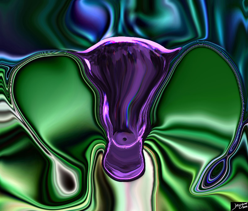
The Flow of the Fallopian Tubes |
| 46701b03.49k.8s uterus fallopian tubes cervix ovaries broad ligament normal anatomy vagina gravid vertex presentation brain placenta foot rawMRI T2 weighted Courtesy Ashley Davidoff MD Davidoff art copyright 2009 all rights reserved |

Normal HSG |
| 30594.8s Normal cervix. Note the serrated appearance of the cavity uterus cervix normal anatomy HSG hysterosalpingogram Courtes Ashley DAvidoff MD copyright 2009 all rights reserved |
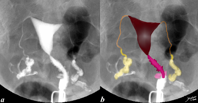
Normal Hysterosalpingogram |
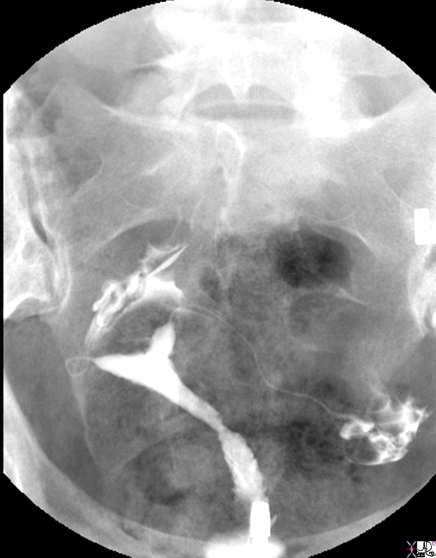
| This hysterosalpingogram is from a 26 year old female with normal endometrial cavity and cervical canal. Note the irregular shape to the cervical canal (dark pink) while the endometrial lining is smooth (marroon). Note also the free spillage of contrast from the tubes (orange) into the peritoneal cavity (white contrast in d,) via the fimbrae (yellow) indicating patent tubes.
code fallopian tubes and fimbrae normal anatomy cervix uterus HSG hysterosalpingogram Courtesy Ashley Davidoff MD Copyright 2009 all rights reserved 83690c02.8s |
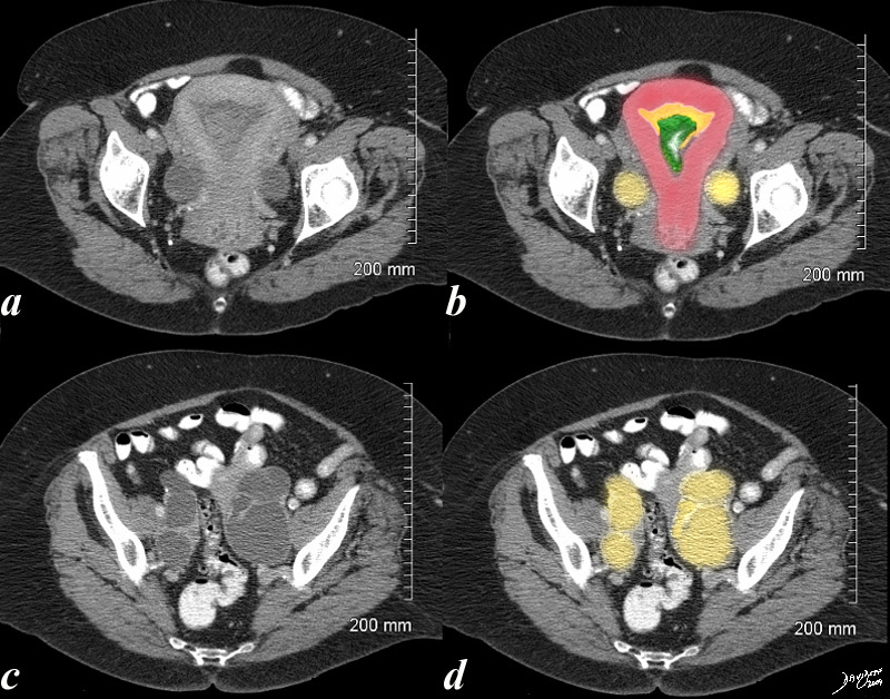
Endometrial Carcinoma with Obstruction of the Uterus and Fallopian Tubes |
| 70 year old female with pelvic discomfort. The CT shows an endometrium filled with complex soft tissue (green), fluid and or blood (orange) and the Fallopian tubes which are distended with fluid (yellow) caused by the obstructing carcinoma.
uterus fallopian tubes endometrial cavity fx mass expanded fx enlarged fx heterogeneous accumulation and expansion of the endometrium fx cytis expansion of the tubes fx enlarged uterus dx endometrial carcinoma with secondary obstruction of the tubes – hydrosalpinx hematosalpinx salpinges CTscan Courtesy Ashley DAvidoff MD 48414c02.8s |
Hysterosalpingogram Fallopian Tubes |
| 46376b01b01b03 pelvis uterus Fallopian tubes fimbrae uterus uterine cavity cervix vagina fornix hysterosalpingogram Davidoff MDpelvis uterus Fallopian tubes fimbrae uterus uterine cavity cervix vagina fornix hysterosalpingogram Davidoff MD |
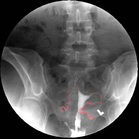
Position and Shape Relates to the Position of the Uterus |
| 40 year old female with normal endometrial cavity and cervical canal and fallopian tubes normal anatomy cervix uterus HSG hysterosalpingogram
Courtesy Ashley Davidoff MD Copyright 2009 all rights reserved 83700.8s |