
Copyright 2010
Definition
Endometrial polyps are benign growths of the endometrium, which occur most commonly in middle-aged women.
The cause of most endometrial polyps is unknown. However, they have been associated with the use of the drug Tamoxifen, as well as hypertension and obesity.
The result is a growth of endometrial glands and stroma projecting from the internal uterine surface. The structural change is characterized by a mass of endometrial glands and fibrous stroma that surrounds a core of large vascular channels. This mass usually arises from the uterine fundus and projects from a stalk into the uterine cavity.
The functional change associated with endometrial polyps may be minimal or limited to abnormal uterine bleeding. However, there is some evidence that removal of endometrial polyps may improve fertility.
Endometrial polyps present clinically with abnormal uterine bleeding. This bleeding is most often light bleeding between periods (metrorrhagia), but can also manifest as heavy or extended bleeding at menses (menorrhagia).
Imaging with ultrasound is useful in the initial investigation of abnormal uterine bleeding, and sonohysterography is the most effective imaging modality for endometrial polyps.
Diagnosis of a benign polyp depends on histologic examination to rule out malignancy.
Treatment of endometrial polyps is surgical excision with hysteroscopy, followed by curettage. Although endometrial polyps are benign, there is a small risk of malignancy arising in a polyp, so removal of large (>1 cm) or symptomatic polyps is recommended.
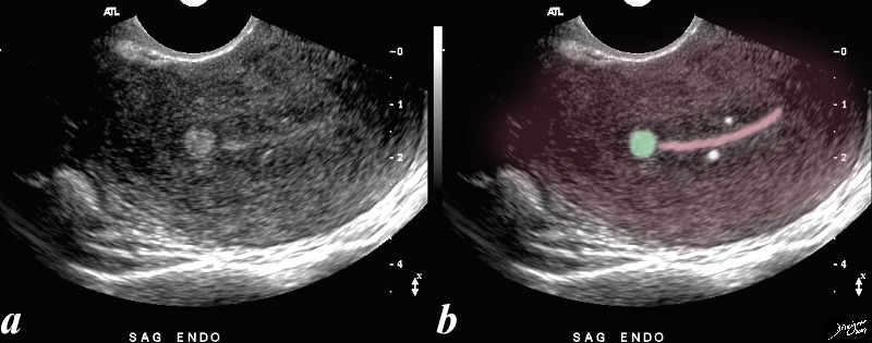
Dystrophic Calcification in Subendometrial Region |
| 52 year-old patient presents with menorrhagia. Two punctate echogenic nodule are in the subendometrial junctional zone, (overlaid in white in b) that are thought to represent dystrophic areas of calcification in prior foci of adenomyosis. The larger echogenic focus (green) was shown to be a benign hyperplastic polyp by pathology after a D and C. The polyp probably accounted for the patient’s menorrhagia. Note that the uterus is retroverted
Courtesy Ashley DAvidoff copyright 2009 all rights reserved 85641b01c.81s |
Submucosal Fibroid |
| The USscan (sonohysterogram) scan is from a 41year old female, who presents with dysfunctional uterine bleeding. The study reveals a 4mms submucosal nodule shown at pathology to be a submucosal fibroid.
Courtesy Ashley Davidoff MD copyright 2009 all rights reserved 84299c041.8 |
|
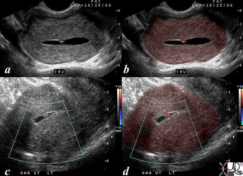
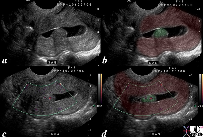
Hyperplastic Polyp |
| The USscan (hysterosonogram is from a 47year old female, with history metrorhagia dysfunctional uterine bleeding. The studies reveal a mass in the uterus within the endometrial cavity The polypwas shown to be a benign hyperplastic polyp.
Courtesy Ashley Davidoff MD copyright 2009 all rights reserved 84299c05.8s |
|
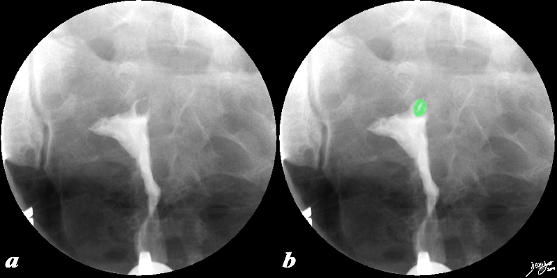
Filling Defect |
|
This 38 year old female presents with a history of infertility. The hysterosalpingogram shows non filling of both Fallopian tubes and a filling defect (green) in the region of the patient’s left cornu that likely is the cause of the obstructed left tube and in part the cause of the infertility. The lesion is most likely a submucosal fibroid but could also be a polyp. Courtesy Ashley Davidoff MD Copyright 2010 All rights reserved 96897c02.8s |
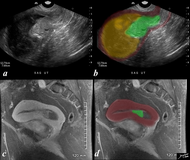
Uterine Polyp with Obstructtion |
| The ultrasound and MRIscan are from a 67 year old female, with an LMP of 10 years ago and with gynecologic history G9 and current history of bleeding for one month, a fever and low hematocrit. She has not had follow up gynecological examination for many years. The studies reveal a mass in the lower uterine segment and an obstructed endometrial cavity tha contains complex material which is presumed combination of blood and pus. ie hematometria and pyometria. The ultrasound is far more impressive than the MRI in that the mass (green) looks more impressive in size and there is prominent upstream accumulation of of complex material and fluids. The MRI shows a enhancing lesion arising from the endometrial mucosa, and at this time the fundal portion is relatively decompressed. The mass is presumably a benign polyp since her subsequent history to the hospital revealed only follow up for chronic renal failure and no pathological report of a malignancy has been n noted.
Courtesy Ashley Davidoff MD copyright 2009 all rights reserved 84127c01.8s |
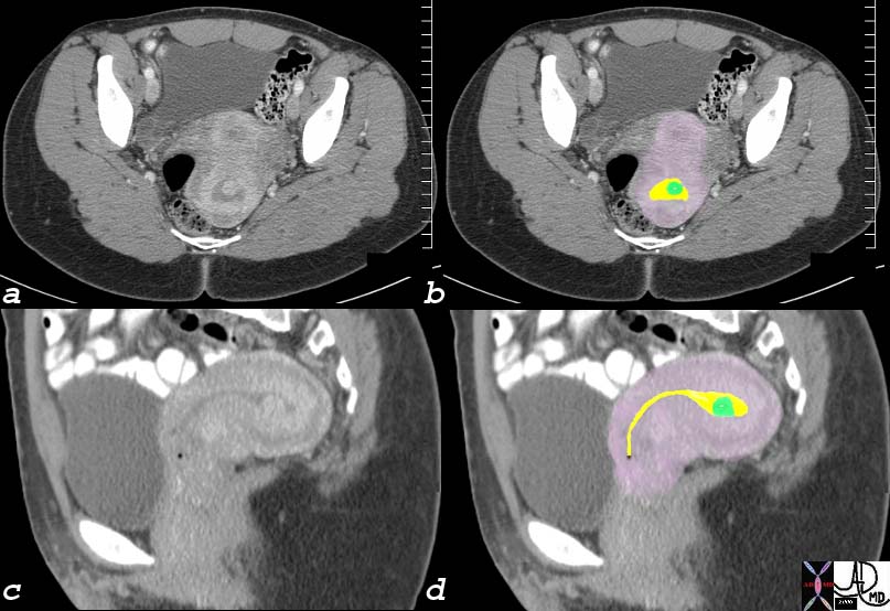
Endometrial Filling Defect – Polyp or Submucosal Fibroid |
|
The coronal reconstruction of a CTscan of a young woman shows a retroverted uterus with a enhancing filling defect (green overlay in b) in the endometrial cavity (yellow). These findings are consistent with either an endometrial polyp or a submucosal fibroid. Endometrial carcinoma is a remote possibility. endometrial fluid = yellow polyp = green myometrium = pink Image Courtesy Ashley Davidoff MD Copyright 2010 76018c01 |