
Principles of the Uterus
The Common vein Copyright 2010
Introduction
The uterus is a muscular pyriform structure specifically designed structurally and physiologically to allow implantation of the embryo and to accommodates a growing fetus until childbirth. The stroma and muscle develop from surrounding mesoderm. The uterus undergoes many changes during the life time of women especially during childbirth. After menopause it becomes atrophic, and may lose its support resulting in prolapse.
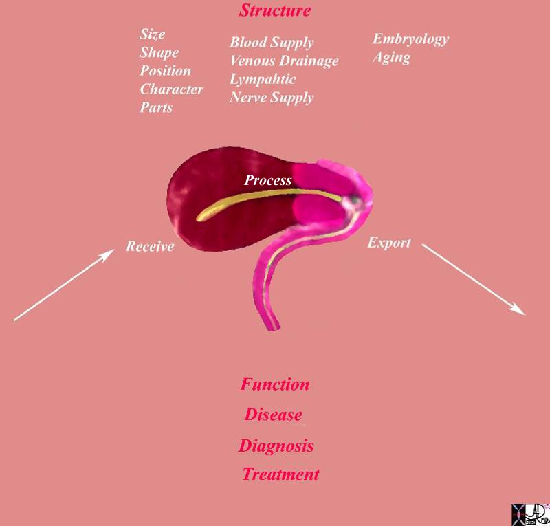
Principles |
|
This diagram frames the underlying principles and approach to the uterus in this module. Image Courtesy Ashley Davidoff MD 93852g.ut01.8s |
As Biological unit:
The uterus is an important reproductive organ and has unique features like its lining of endometrium that changes with menstrual cycle to allow implantation of the fertilized embryo and cris-cross myometrium that stretches to accommodate a growing fetus. The cris-cross myometrium also acts as living ligatures to minimize blood loss during and after child birth. The fallopian tubes acts as a conduit for the unfertilized ovum to to be transported to the endometrial cavity to enable fertilization.
Links and Connections
The uterus is connected to the fallopian tubes superiorly and to the vagina inferiorly. It is a pelvic organ and lies between urinary bladder anteriorly and rectum posteriorly. It has an independent blood and lymphatic supply. It has unique ligaments and a muscular sling that helps in maintaining its position and prevent uterine prolapse. It is intimately comnnected to the endocrine system via circulating hormones particularly estrogen and progesterone.
Time growth and Aging
During organogenesis a pair of Mullerian ducts arise from coelomic epithelium and subsequently fuse to form the uterus. The proximal portions form the fallopian tubes and distal portions forms the body and cervix of uterus and upper 4/5ths of the vagina
Dependence and independence
The uterus along with ovaries forms the female reproductive system and is an independent unit. However, the functioning of uterus is under a complicated system of hormones secreted by ovaries which in turn is controlled by pituitary hormones. Some pituitary hormones also have a direct effect on uterus. This constitutes a pituitary-ovarian – uterine axis.
Spaces
The non gravid uterus is a pelvic organ. However a gravid uterus becomes an abdominal organ occupying a large area in abdomen almost up to the tip of the xiphisternum at term. It is also only partially covered by peritoneum on its superior aspect.
Forces
Uterine contractions can occur in the non pregnant state. During the menses, contractions occur to help eject the shedded endometrium. During pregnancy the uterus undergoes painless contraction called Braxton Hicks which increase as the pregnancy advances. During parturition uterine contractions have two separate effects in the upper uterine segments. Thereis one set of contractions which gradually create a downward force and shortening of the muscle fibres which tend to get smaller by a process called retraction, causing descent of the fetus and rotation of the fetal head. The second effect is on the cervix and lower uterine segment which dilates as upper uterine segment contracts.
Interactions
The uterus, ovaries and pituitary form a sophisticated unit playing an important part in ovulation, conception and child birth. The uterus along with the fallopian tubes and cervix is structural pillar of this unit.
State of health and disease
Uterine problems are very common manifesting as fibroid disease, dysfunctional uterine bleeding, structural anomalies and uterovaginal prolapse.
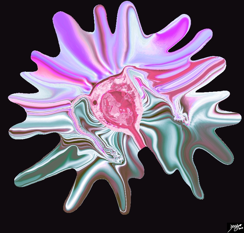
The Uterus – Our Survival Depends on It |
|
Life depends on the uterus. The uterus is the ultimate organ that enables the survival of the human race. It houses, protects and nourishes the fetus for approximately 9months and bears its fruit by delivering its product through the use of strong muscular forces. Courtesy Ashley Davidoff MD Copyright 2010 all rights reserved 46701b03.48kd13.8s |
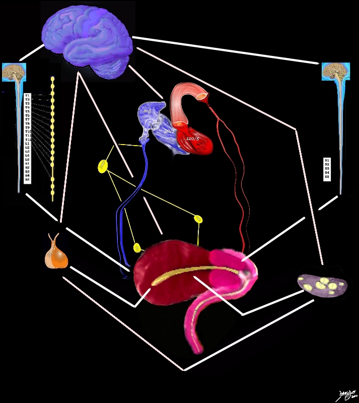
Integrated Function |
|
The image shows some of the structures in the body that are functionally and structurally linked to the uterus in order for it to functions as an integrated organ. The brain (purple reigns) supreme and is responsible for the neural axis, the pituitary (orange) and ovary (black and gray) for the hormonal axis. The vascular axis is exemplified by the arteries and veins (red and blue) while the lymphatic connections are outlined in yellow Courtesy Ashley Davidoff MD copyright 2010 all rights reserved 46701b03.48ke05.86s |